Quick Facts
A venule is any of the small vessels that collect blood from the capillary plexuses and join to form veins (Dorland, 2011).
Structure
Blood vessels are tubular structures in which blood circulates around the body, creating a network which, along with the heart, we refer to as the circulatory system or cardiovascular system.
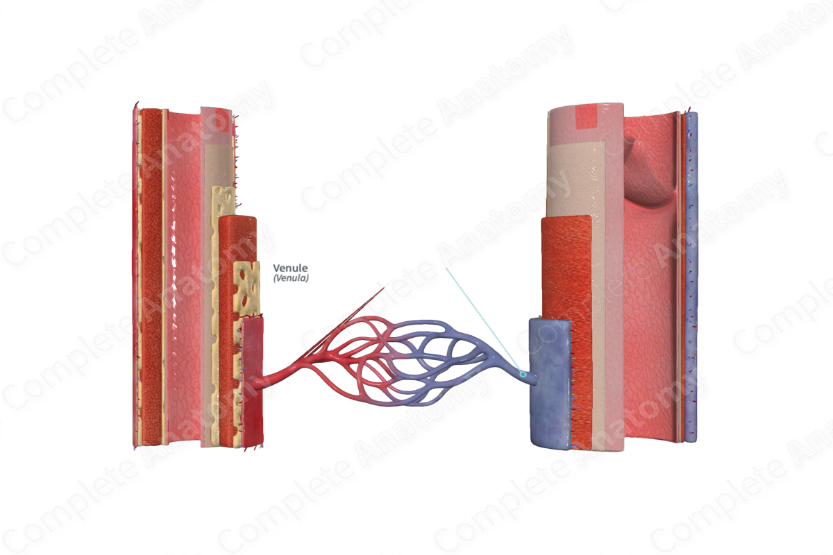
Venules are vessels which lie between capillaries and veins, thus connecting them together. They have extremely thin walls, similar to that of capillaries, which give them a misshapen or flattened appearance in a cross-sectional view.
Veins have three principal layers in their wall; the innermost tunica intima, surrounded by a middle layer called the tunica media, and a superficial layer called the tunica externa. The structure of the walls of venules differs from this composition (explained below).
There are two types of venules.
- Postcapillary venules, which are found directly after capillary beds, are typically 10-30μm in diameter, only slightly larger than that of capillaries (Standring, 2016). Postcapillary venules are composed of an endothelial layer and a basal lamina surrounded by a thin connective tissue layer in place of the tunica externa.
- True venules, also known as muscular venules, are typically distinguished from postcapillary venules where their diameter exceeds 50μm and smooth muscle cells are found in their walls (Standring, 2016).
Function
All venous vessels carry blood from body organs and tissues back to the heart. They typically transport deoxygenated blood, with the exception of the venules within the pulmonary circulation. These vessels are involved in the transportation oxygenated blood from the lungs to the heart.
As the postcapillary venules have a composition similar to that of capillaries, they may be involved in gas and solute exchange with surrounding tissues. The movement of leukocytes into extravascular spaces, also known as leukocyte migration, also occurs in postcapillary venules (Standring, 2016).
Muscular venules are not involved in gas and solute exchange due to their more substantial wall thickness. Muscular venules will eventually merge and form veins, which will continue to carry blood back towards the heart.
Because of the low levels of elastin fibers, lower mass of smooth muscle (in comparison to arteries), and relatively large lumen, venules and veins can be stretched and expanded (distensible). The distensibility of veins enables them to act as a blood reservoir, allowing storage of large volumes of blood. This can occur when blood returning to the heart is disrupted due to low pressure or compression.
List of Clinical Correlates
- Venous insufficiency
- Deep vein thrombosis
- Portal hypertension
- Varicose veins
References
Dorland, W. (2011) Dorland's Illustrated Medical Dictionary. 32nd edn. Philadelphia, USA: Elsevier Saunders.
Standring, S. (2016) Gray's Anatomy: The Anatomical Basis of Clinical Practice. Gray's Anatomy Series: Elsevier Limited.
