Quick Facts
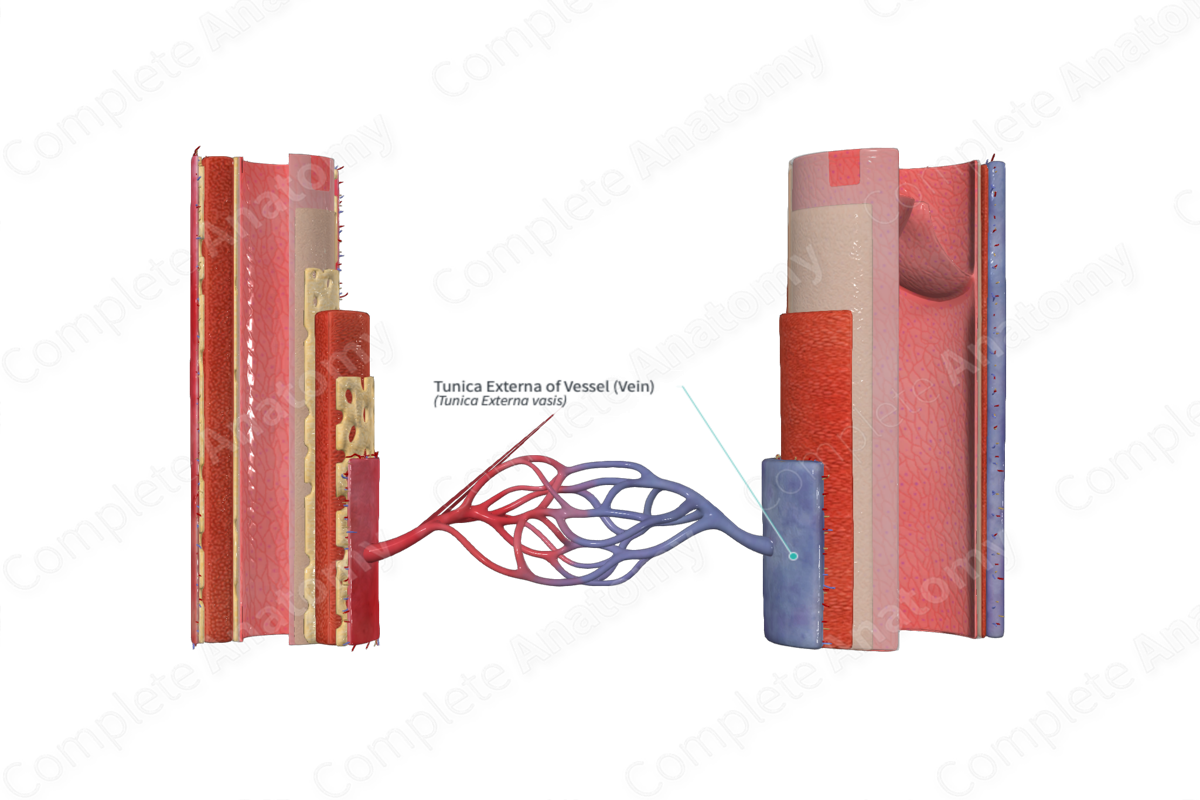
The tunica externa of a blood vessel is the outer, fibroelastic coat of vessels, usually in contact with surrounding tissues (Dorland, 2011).
Structure
The tunica externa is the outer layer of the vessel wall, in veins this is the most prominent of the three main layers.
The tunica externa is composed of connective tissue, elastin and type I collagenous fibers, nerves and the network of vessel capillaries, the vasa vasorum. The tunica externa of large blood vessels also contains a network of unmyelinated autonomic nerve fibers, called nervi vasorum (Mescher, 2013).
Recent studies have suggested that the tissue arrangement of tunica externa is not a densely packed wall like-structure, commonly seen in alcohol fixed tissue. In fact, it is composed of a collagen lattice surrounded by fluid filled interstitial spaces (Benias et al., 2018). This has also been described in other tissues, such as lung, renal, dermis, and fascia.
Anatomical Relations
The tunica externa is the outermost layer of the blood vessel. It is loosely formed, continuous and covers the entire outer portion of the blood vessel serving to anchor the blood vessel in place in the body. It lies on top of the tunica intima separated by the external elastic lamina.
Function
The tunica externa acts as an anchor, securing the blood vessel to the surrounding body tissue.
References
Benias, P. C., Wells, R. G., Sackey-Aboagye, B., Klavan, H., Reidy, J., Buonocore, D., Miranda, M., Kornacki, S., Wayne, M., Carr-Locke, D. L. and Theise, N. D. (2018) 'Structure and Distribution of an Unrecognized Interstitium in Human Tissues', Scientific Reports, 8(1), pp. 4947.
Dorland, W. (2011) Dorland's Illustrated Medical Dictionary. 32nd edn. Philadelphia, USA: Elsevier Saunders.
Mescher, A. (2013) Junqueira's Basic Histology: Text and Atlas. 13th edn.: McGraw-Hill Education.
