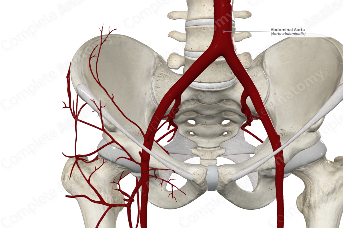
Quick Facts
Origin: Continuation of the thoracic aorta at the level of T12.
Course: Courses inferiorly as far as L4.
Branches: Inferior phrenic arteries, celiac trunk, superior mesenteric, middle suprarenal, renal, gonadal (testicular or ovarian), inferior mesenteric, lumbar, median sacral, and common iliac arteries.
Supplied Structures: Abdominal viscera, abdominal wall, and diaphragm.
Origin
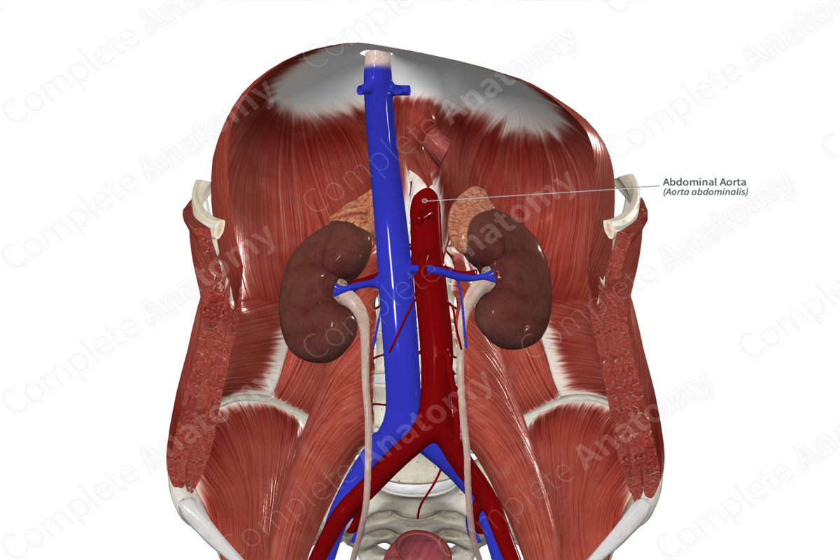
The abdominal aorta originates as a continuation of the thoracic aorta as it passes through the aortic hiatus of the respiratory diaphragm at the level of T12 vertebra.
Course
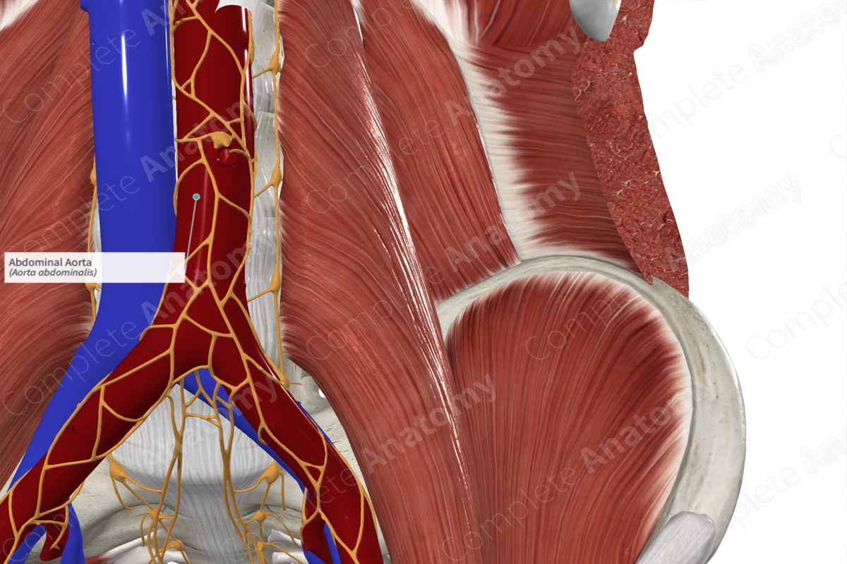
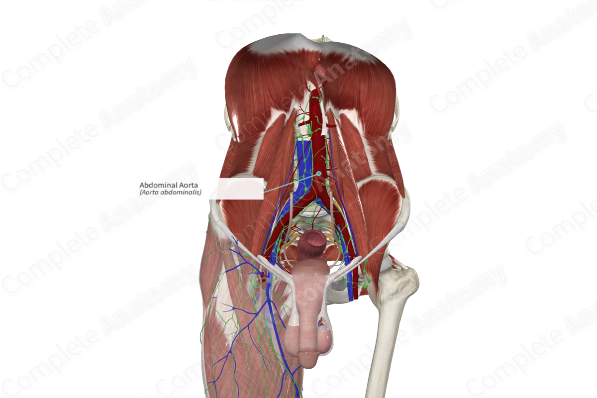
It travels inferiorly, slightly to the left of the midline, anterior to the vertebral column. At the level of L4 vertebra, it bifurcates into the right and left common iliac arteries.
Branches
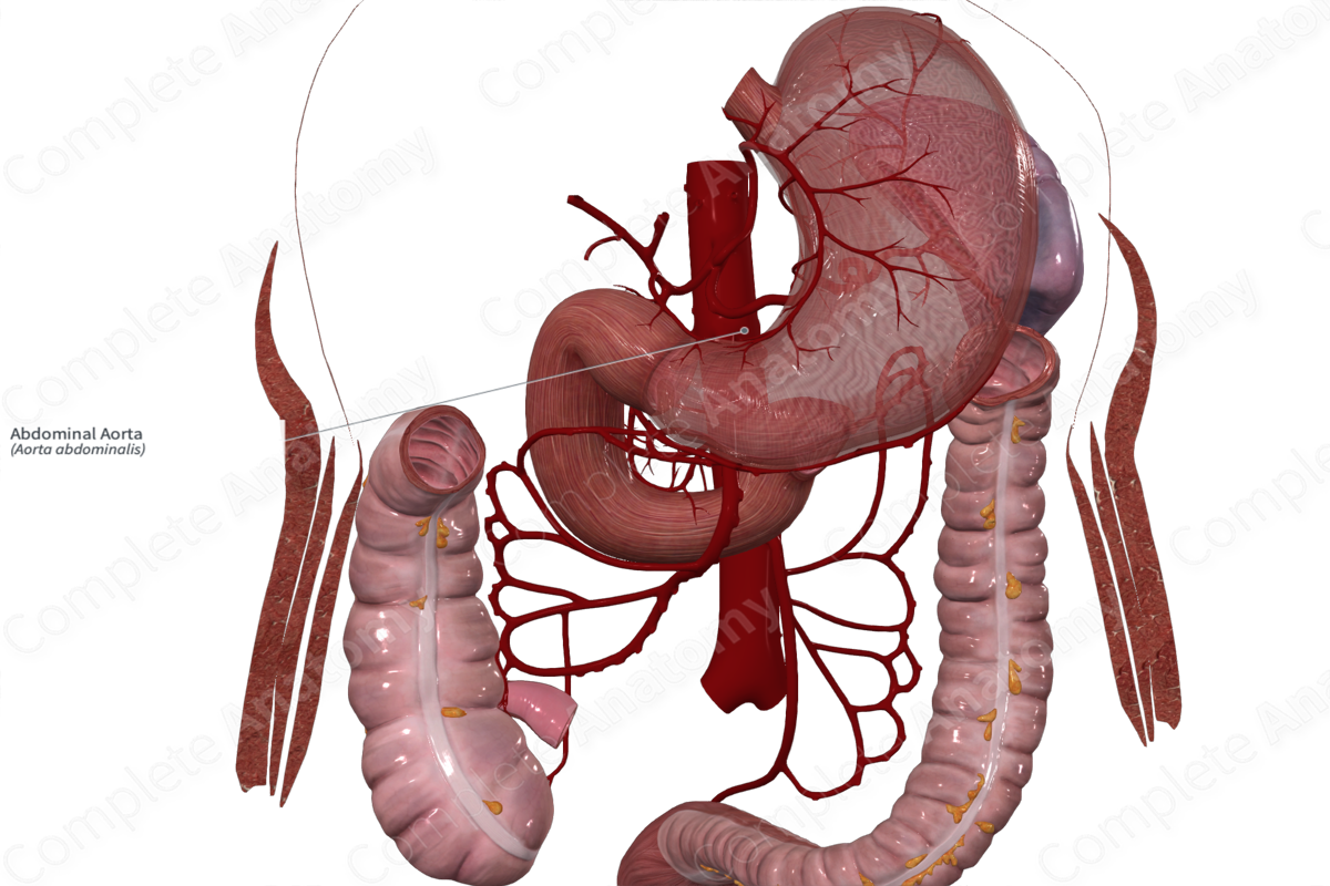
The branches of the aorta can be divided into three groups, including unpaired visceral, paired visceral, and parietal branches. The unpaired visceral branches arise from the anterior aspect of the abdominal aorta and supply the organs of digestion and the spleen. These branches include (in descending order) the celiac trunk, the superior mesenteric artery, and the inferior mesenteric artery.
The next group of branches are the paired visceral branches. The three paired branches arise from the abdominal aorta between the superior mesenteric artery and inferior mesenteric artery. They are the middle suprarenal arteries, the renal arteries, and the gonadal (ovarian or testicular) arteries.
The last group of branches are small branches which mainly supply the abdominal walls and the diaphragm. They include the inferior phrenic arteries, the lumbar arteries, and the median sacral artery.
The terminal branches of the abdominal aorta are the right and left common iliac arteries.
Supplied Structures
The three unpaired visceral branches (the celiac trunk, superior mesenteric artery, and inferior mesenteric artery) supply the foregut, midgut, and hindgut, respectively.
The paired visceral branches supply the suprarenal glands, kidneys, and testes or ovaries.
The inferior phrenic arteries contribute to the diaphragmatic supply. The lumbar arteries supply the posterior abdominal wall, while the median sacral artery supplies the sacrum and coccyx.
The common iliac arteries further divide and supply the pelvis, perineum, and lower limbs.
List of Clinical Correlates
- Aortic aneurism
Learn more about this topic from other Elsevier products

