Quick Facts
Origin: Proximal portion of the ascending aorta.
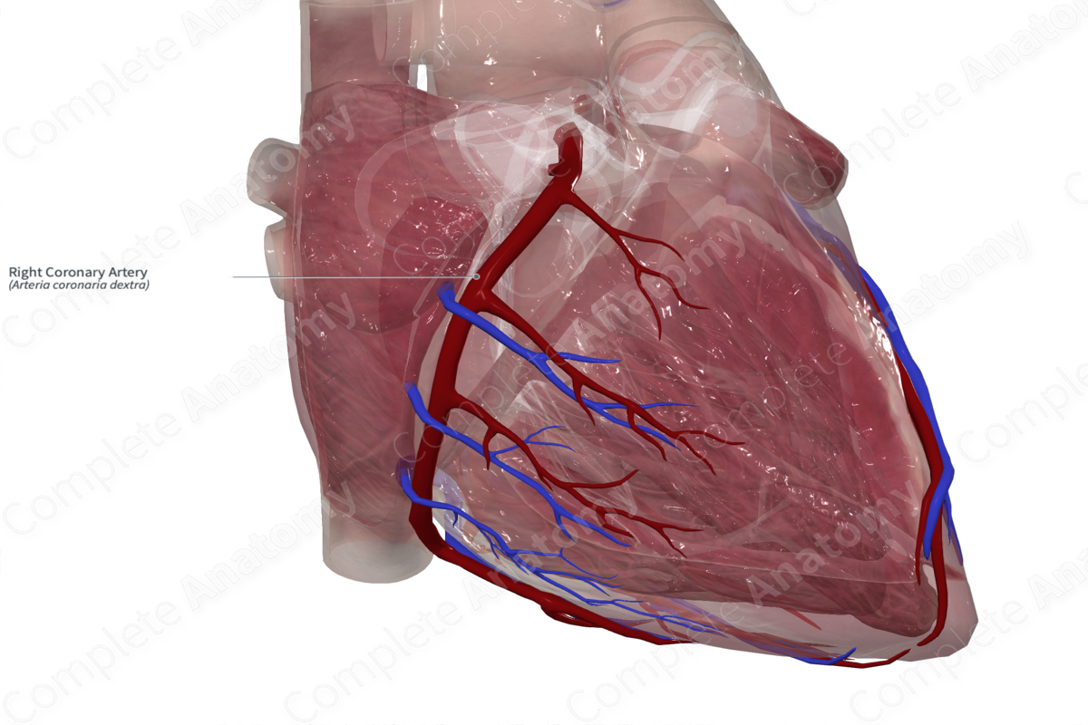
Course: It runs in the coronary sulcus from the anterior aspect heart to the posterior interventricular sulcus.
Branches: Conus, atrial, and ventricular branches; right marginal arteries; atrioventricular nodal, sinuatrial nodal, posterior interventricular, and right posterolateral branches.
Supplied Structures: Right atrium, most of right ventricle, a portion of the left ventricle (inferior surface), posterior one third of the interventricular septum, sinuatrial node (in approx. 60% of individuals) and the atrioventricular node (in approx. 80% of individuals).


Related parts of the anatomy
Origin
The coronary arteries are the first branches of the thoracic aorta and the only branches of the ascending aorta, arising from its proximal portion below the sinutubular junction.
The right coronary artery arises from the right coronary aortic sinus. It is smaller in diameter than the left coronary artery. The arteries are subepicardial but may be more deeply embedded in regions such as the interventricular grooves. In addition, they are covered by adipose tissue and may be somewhat hidden on initial inspection of the heart during cadaveric dissection.
Course
The right coronary artery first courses in an anterior direction, then towards the right where it sits between the right atrial appendage and the pulmonary trunk to reach the coronary sulcus (atrioventricular groove). In this groove, it descends towards the right in a near vertical manner, along the right cardiac border. It then curves posteriorly where it reaches the junction between the interatrial and the interventricular grooves (the cardiac crux).
The position of the termination of the artery is variable. It often terminates to the left of the crux where it frequently joins the circumflex branch of the left coronary artery (Standring, 2016).
Branches
The conus branch is the first branch of the right coronary artery. There are a series of small branches that supply the anterior aspect of the right atrium and ventricle. Of these branches there are usually two or three anterior ventricular branches.
The next branch is the right marginal artery. It is the largest of the ventricular branches and runs inferiorly where it almost reaches the apex. There are also a series of posterior ventricular branches. It then gives the posterior interventricular artery and often the right posterolateral branch of the right coronary artery.
The right coronary artery usually gives off both the sinuatrial nodal branch and the atrioventricular nodal branch (Standring, 2016).
Supplied Structures
The right coronary artery supplies almost the entire right ventricle, as well as the right atrium and a portion of the left atrium. It supplies the posterior one third of the interventricular septum. It also contributes to the supply of the conduction system, including the sinuatrial node and the atrioventricular node.
The arterial supply to the diaphragmatic surface of the heart is the most variable in terms of right or left coronary arterial supply. Its supply is related to which vessel is dominant. The term dominant does not refer to the amount of cardiac tissue supplied by the arteries, but from which coronary artery that gives rise to the inferior interventricular artery (aka the posterior descending artery [PDA] or posterior interventricular artery). This artery supplies the diaphragmatic inferolateral wall of the left ventricle. The right coronary artery is dominant in 67% of individuals (Moore, Dalley and Agur, 2013).
List of Clinical Correlates
- Coronary artery disease
- Coronary atherosclerosis
- Coronary bypass graft
- Coronary angioplasty
- Coronary occlusion
- Coronary revascularization
- Coronary artery fistula
References
Moore, K. L., Dalley, A. F. and Agur, A. M. R. (2013) Clinically Oriented Anatomy. Clinically Oriented Anatomy 7th edn.: Wolters Kluwer Health/Lippincott Williams & Wilkins.
Standring, S. (2016) Gray's Anatomy: The Anatomical Basis of Clinical Practice. Gray's Anatomy Series 41st edn.: Elsevier Limited.
Learn more about this topic from other Elsevier products

