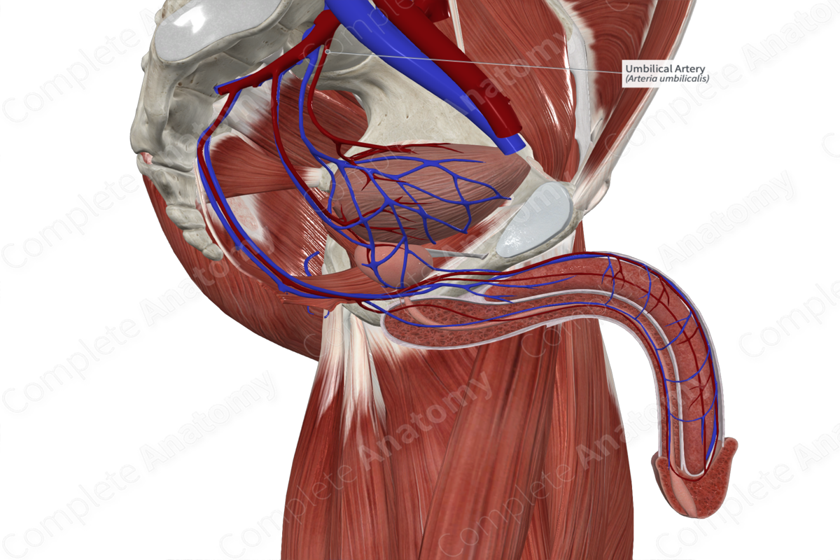
Quick Facts
Origin: Internal iliac artery.
Course: Travels anteriorly to abdominal wall, then superiorly to the umbilicus.
Branches: Superior vesical arteries and the artery to ductus deferens.
Supplied Structures: Superior bladder, inferior ureter, ductus deferens, and seminal vesicle.
Origin
Paired umbilical arteries arise from the anterior division of the internal iliac artery. It is usually an early proximal branch.
Course
The umbilical artery passes anteromedially across the pelvis to the anterior abdominal wall. It then passes superiorly, along the inner surface of the anterior wall. Right and left arteries pass medially as they ascend to converge on the umbilicus. In the fetus, this vessel is entirely patent and passes through the abdominal wall at the umbilicus, into the umbilical cord to the placenta. After birth the vessel becomes fibrotic, except for the most proximal, pelvic portion that gives off the superior vesical arteries. The occluded part of the umbilical artery forms the medial umbilical folds, which are located along the internal surface of the anterior abdominal wall.
Branches
Up to four superior vesical arteries arise from the proximal part of the umbilical artery.
In males, the artery may give off a branch to the ductus deferens. However, there is considerable anatomical variation in the origin of this vessel. The artery to the ductus deferens may branch exclusively from the superior vesical arteries.
Supplied Structures
The umbilical artery supplies the superior portion of the bladder. In males, the artery gives off variable branches that supply the ductus deferens, testis and seminal vesicle.
Learn more about this topic from other Elsevier products


