Quick Facts
Origin: Subclavian artery.
Course: Ascends through the foramina transversaria of the C1–C6 vertebrae and the foramen magnum to enter the cranial cavity.
Branches: Basilar, anterior spinal, posterior inferior cerebellar, and meningeal arteries, spinal and muscular branches.
Supplied Structures: Muscles of the neck, vertebrae, spinal cord, cerebellum.
Related parts of the anatomy
Origin
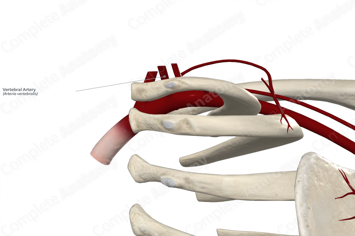
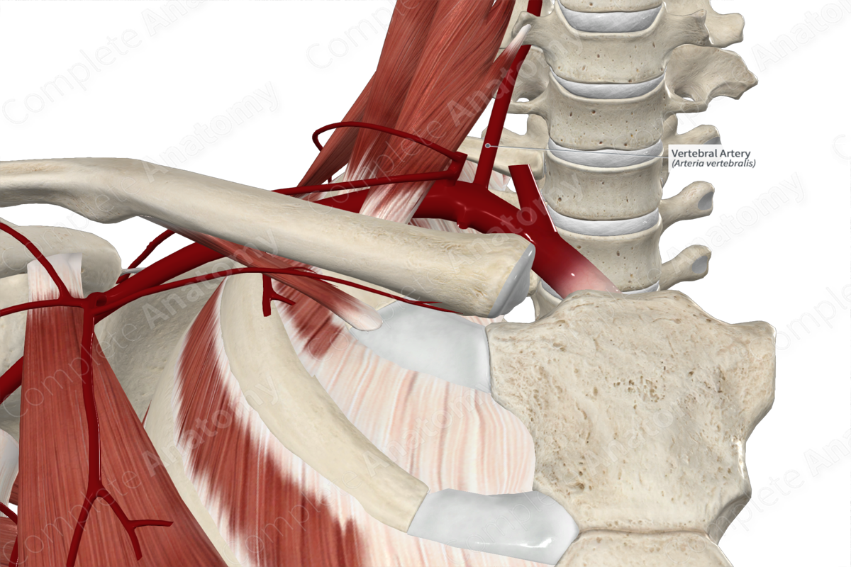
The vertebral artery originates from the posterosuperior aspect of the subclavian artery, close to the lateral aspect of the vertebral bodies. In 5% of people, the left vertebral artery arises from the aorta (Moore, Dalley and Agur, 2013).
Course
For descriptive purposes, the course of the artery can be divided into four parts; the cervical, vertebral, suboccipital, and cranial parts.
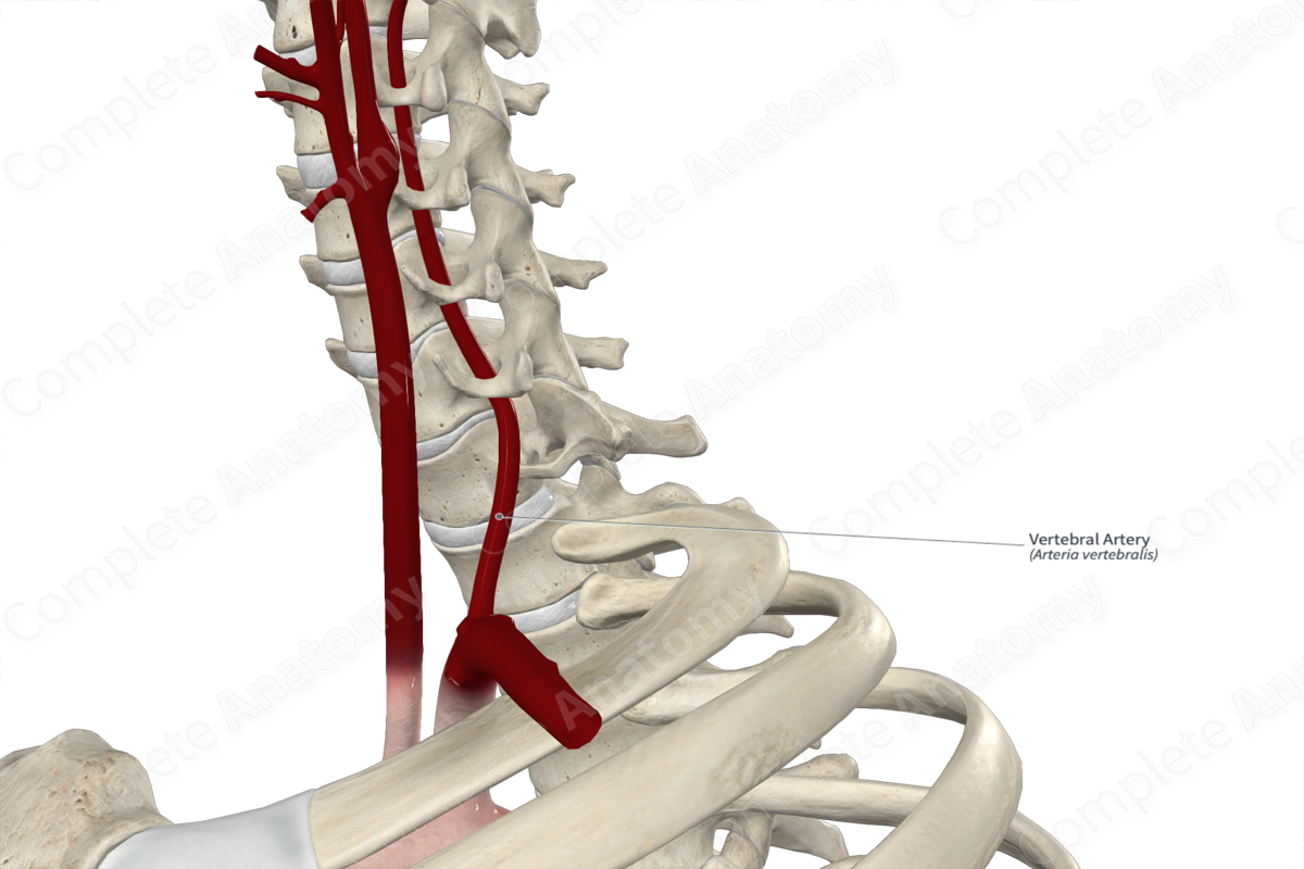
From its origin, the cervical part of the vertebral artery travels superiorly between the longus colli and scalenus anterior muscles.
The vertebral part then passes through the foramina transversaria of the sixth cervical vertebra. It continues to ascend towards the cranium by traveling through the remaining cervical foramina transversaria (C5–C1).
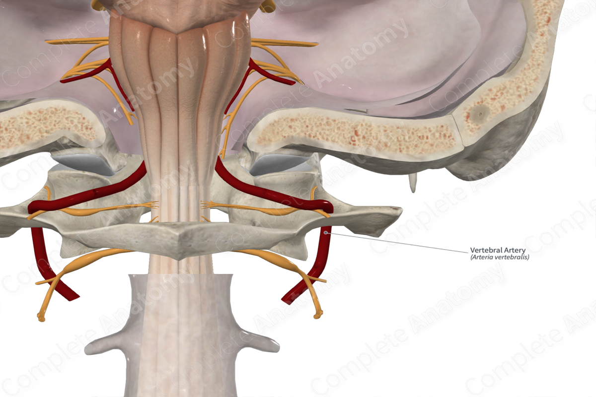
After the vertebral artery passes through the foramen transversarium of the atlas, the suboccipital part of the vertebral artery travels in a groove on the superior portion of the posterior arch of the bone. The artery then ascends through the foramen magnum to enter the cranial cavity.
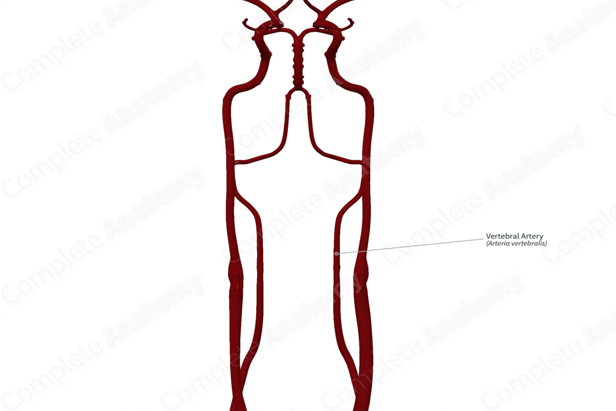
The cranial part of the vertebral artery takes a superomedial course on the surface of the medulla. It converges with the vertebral artery of the opposite side at the junction between the medulla and pons to form the basilar artery.
Branches
The vertebral artery and its branches are sometimes referred to as the vertebrobasilar system. Close to the foramen magnum, meningeal branches arise from the vertebral artery. Near the union of the vertebral arteries, the anterior spinal artery arises and descends on the spinal cord. The posterior inferior cerebellar artery is sometimes absent, but when present it arises from the vertebral artery close to the olive.
Supplied Structures
The vertebral artery supplies the upper spinal cord and cerebellum.
References
Moore, K. L., Dalley, A. F. and Agur, A. M. R. (2013) Clinically Oriented Anatomy. Clinically Oriented Anatomy 7th edn.: Wolters Kluwer Health/Lippincott Williams & Wilkins.
Learn more about this topic from other Elsevier products

