Morphology/Structure
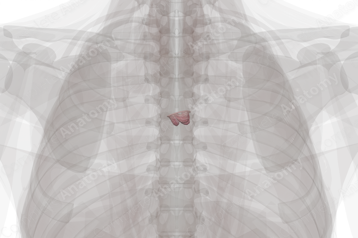
The aortic valve is located at the aortic root, which bridges the vestibular outflow tract of the left ventricle and the aorta. The aortic valve is composed of a fibrous aortic ring and three semilunar leaflets (or semilunar cusps); the right, left, and noncoronary (nonadjacent) semilunar leaflets.
The leaflets are covered with endocardium and are attached to the aortic wall and supporting structures of the ventricles. The semilunar shape of the leaflets ensures that the leaflets are in close proximity to each other during diastole. To aid in apposition, the free edges of the leaflets are thickened in a region called the lunule, the apex of which is called the nodule.


Related parts of the anatomy
Key Features/Anatomical Relations
The leaflets are supported within the sinuses (of Valsava). The sinuses extend from their proximal attachment beyond the free margin of the semilunar leaflets to the sinutubular junction. This is the junction between the aortic sinus and the ascending aorta.
The location of the fibrous rings is often described as the origin of the sinuses where the semilunar hinges of the valve leaflets attach. The coronary arteries open into the upper portions of the sinuses. The right semilunar leaflet lies next to the ostia of the right coronary artery and is therefore also known as the right coronary leaflet. The left semilunar leaflet lies adjacent to the ostia of the left coronary artery and is therefore also known as the left coronary leaflet. The aortic sinuses encourage non-turbulent blood flow through the openings of the coronary vessels.
The semilunar valves do not require chordae tendineae or papillary muscles to function, as their semilunar attachment to the wall of the pulmonary trunk gives them their inherent stability. When the valve is closed, the commissures are the points of apposition between adjacent leaflets. The degree of apposition that is possible is essential to the determination of valvular competence.
The aortic valve can be auscultated in the second intercostal space, along the right sternal edge, below the manubriosternal joint.
Function
The aortic valve permits unidirectional flow of oxygenated blood from the left ventricle to the aorta and prevents retrograde flow. When the heart is filling during diastole, the blood in the aorta is diminishing in pressure thus each sinus and their leaflets are opposed, and the valve is closed.
When ventricular pressure increases during systole, the valve is passively open, and blood is ejected from the ventricle. During the ejection of blood, some blood pools in the aortic sinuses and allows it to enter the coronary vessels. The valve then closes again in diastole.
List of Clinical Correlates
- Aortic stenosis
- Aortic regurgitation
- Infective endocarditis
- Rheumatic heart disease
- Marfan’s syndrome
Learn more about this topic from other Elsevier products

