Description
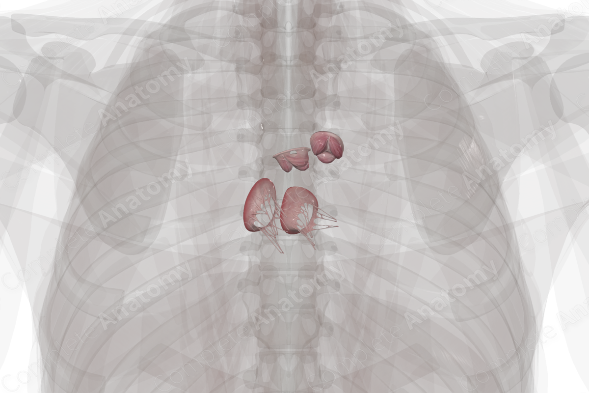
The heart has four valves: two atrioventricular valves and two semilunar valves. All four valves have leaflets which open and close in a fashion that ensures unidirectional blood flow.
The right and left atrioventricular valves are also known as the tricuspid and mitral valves, respectively. These valves require chordae tendineae and papillary muscles to ensure their closure during the ejection of blood from the ventricles.
The two semilunar valves sit between the ventricular outflow tracts and the major arteries. The aortic valve sits at the base of the ascending aorta, while the pulmonary valve sits at the beginning of the pulmonary trunk. These valves do not require the same extensions as the atrioventricular valves and rely on their shape and stability to ensure unidirectional blood flow.
There is a discrepancy in the nomenclature used to describe the components of the valves. The moving portions of the valve are the leaflets. However, for the semilunar valves, they may be referred to as cusps due to their shape. Occasionally they’re incorrectly called sinuses. The sinuses are in fact the arterial dilations that extend superior to the leaflets. Additionally, the term anulus is somewhat inaccurate as they’re not fully ring-shaped and are often discontinuous structures.


Related parts of the anatomy
