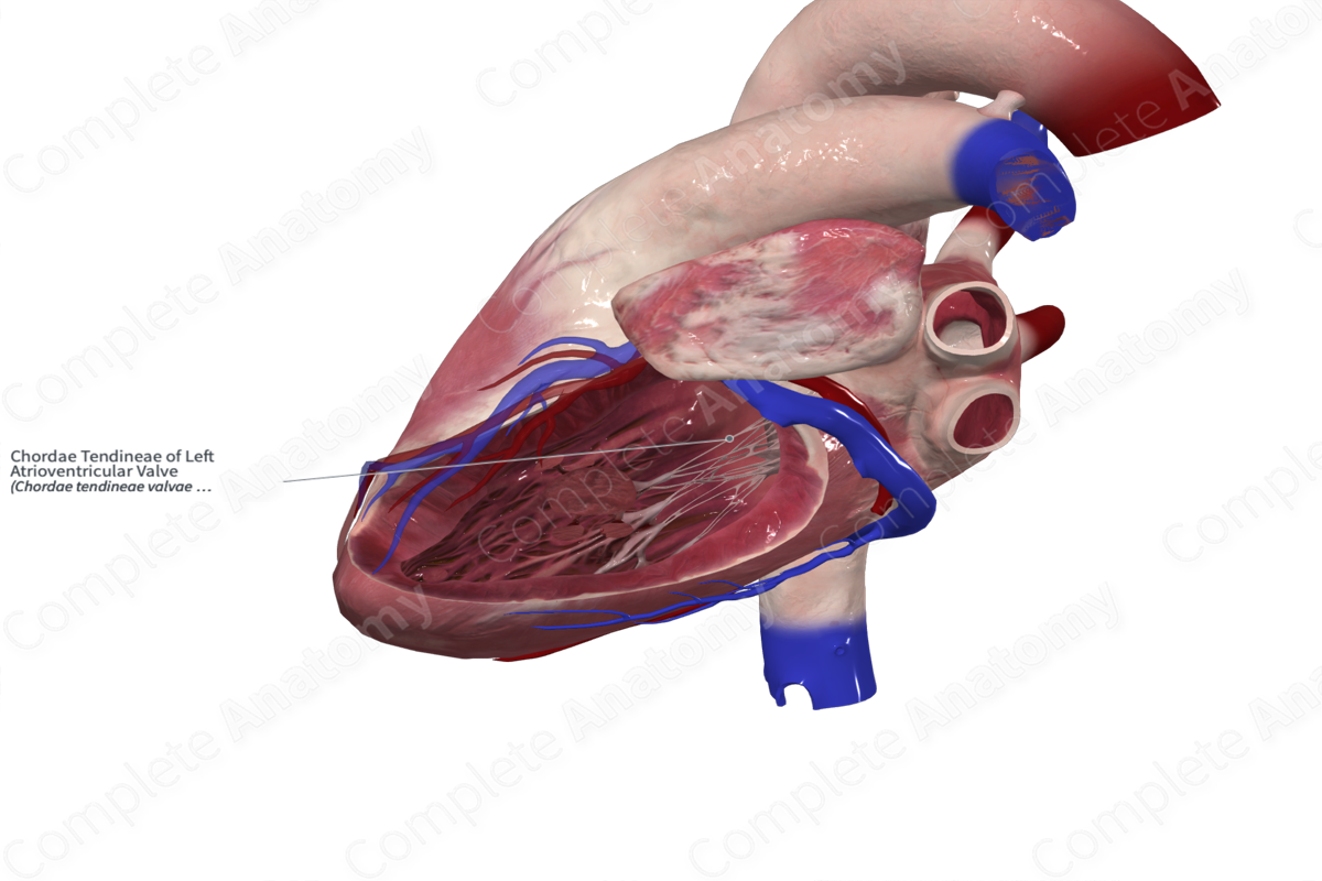
Chordae Tendineae of Left Atrioventricular Valve
Chordae tendineae valvae atrioventricularis sinistrae
Read moreMorphology/Structure
The chordae tendineae of the left atrioventricular valve are slender fibrocollagenous bands that connect the free margins of the atrioventricular valve leaflets to the apical one third of the papillary muscles. The rough zone of each leaflet contains a roughened free margin which provides an attachment site for the bulk of the chordae tendineae. The chordae tendineae of the left ventricle are like those found in the right ventricle.


Key Features/Anatomical Relations
There are several types of true chordae tendineae, including rough and basal zone chordae, which refers to the portion of the leaflet that they attach to. The majority of the chordae tendineae attach to the apical one third of the papillary muscles.
Function
The chordae support the free edges of the leaflets and prevent them from prolapsing when blood is being ejected from the ventricle. During atrial contraction, the papillary muscles are relaxed, and the valve is open, which avoids resistance to the movement of blood from the atrium to the ventricle. During ventricular contraction, blood moving towards the atrium pulls the valve leaflets closed, increasing the tension in the papillary muscles, and thus the chordae tendineae. This tension prevents the leaflets swinging backwards into the atria during ejection of blood from the ventricles. The tension in the papillary muscles remains until the next atrial contraction.
Learn more about this topic from other Elsevier products
Atrioventricular Node

The AV node is a small subendocardial structure located within the interatrial septum at the distal convergence of the preferential internodal conduction pathways that course through the atria from the sinus node.
