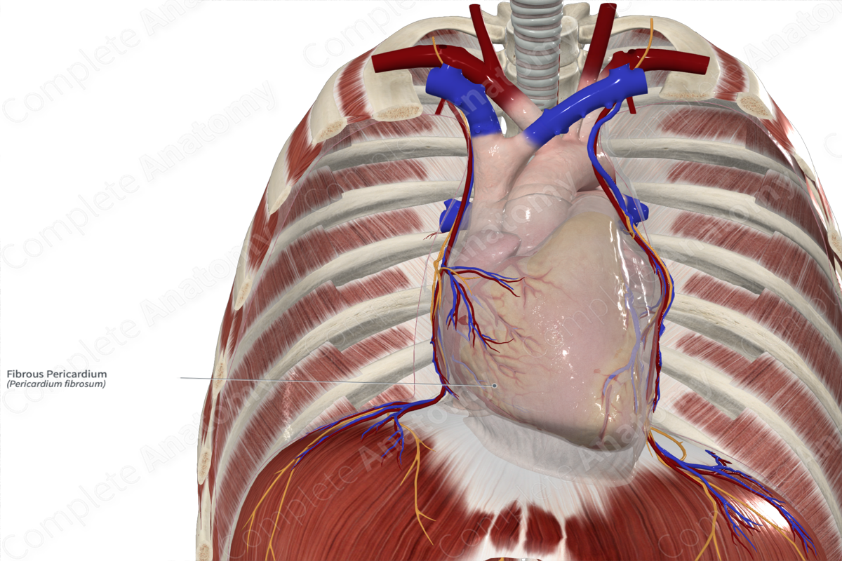
Morphology/Structure
The pericardium envelops the heart and the base of the great vessels. It consists of an outer fibrous layer and two inner serous pericardial layers. The fibrous layer is composed of a three layers of collagen fibers orientated obliquely to each other. This inhibits stretch and provides a barrier to disease.
Key Features/Anatomical Relations
The fibrous layer is attached to:
- the sternum anteriorly via the superior and inferior sternopericardial ligaments;
- the central tendon of the diaphragm inferiorly;
- the vertebral column posteriorly;
- and is continuous with the lungs and mediastinal pleural laterally and the outer layer of the base of the great vessels.
The fibrous pericardium is not attached to the heart itself.
The phrenic nerve and the pericardiacophrenic vessels are enveloped within the fibrous pericardium and run inferiorly along the lateral aspects of the pericardial sac as they travel towards the diaphragm. The arteries and veins provide the bulk of the vasculature of the pericardium, while the phrenic nerves provide sensory innervation to the fibrous pericardium (and the parietal layer of serous pericardium).
Function
The fibrous layer ensures stability by attaching to surrounding structures namely the sternum, vertebral column, and diaphragm. It also provides a physical limit to prevent excessive dilation or overfilling in certain types of heart failure. Additionally, it provides a barrier to the spread of infection.
List of Clinical Correlates
- Cardiac tamponade
- Pericardial effusion
- Pericarditis
- Pericardial rub
Learn more about this topic from other Elsevier products


