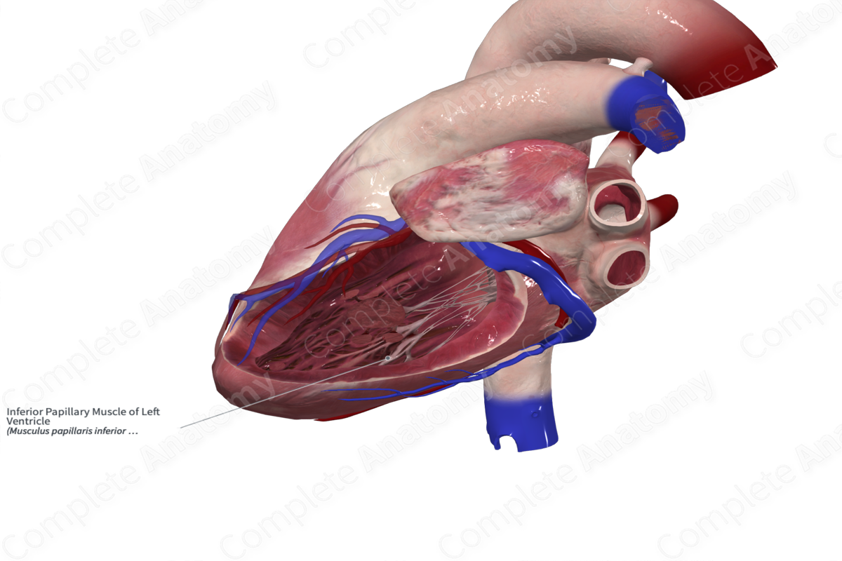
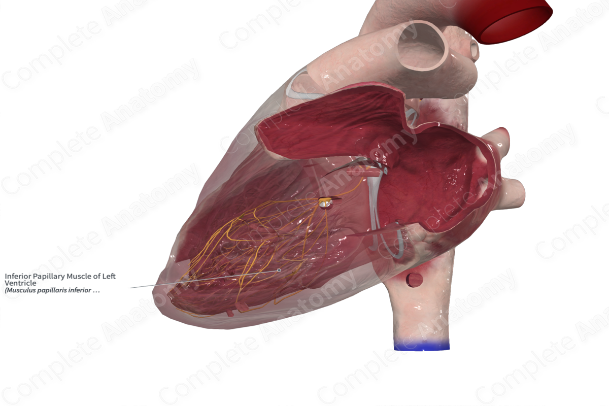
Inferior Papillary Muscle of Left Ventricle
Musculus papillaris inferior ventriculi sinistri
Read moreMorphology/Structure
The papillary muscles are conical muscular bundles that extend from the ventricular wall to the chordae tendineae. The chordae tendineae in turn insert into the leaflets of the left atrioventricular valve (or mitral valve). The chordae tendineae attach to the apical one third of the papillary muscles, but occasionally, attach to the base of the papillary muscles.


Key Features/Anatomical Relations
There are two papillary muscles in the left ventricle. They vary in dimensions and are often bifid. The inferior papillary muscle arises from the inferior (posterior) ventricular wall. The superior papillary muscle arises from the anterior ventricular wall. The papillary muscles attach to the aortic (anterior cusp) and mural (posterior cusp) leaflets of the left atrioventricular valve to form a single commissural region.
Function
During atrial contraction, the papillary muscles are relaxed, and the valve is open, avoiding any resistance to the movement of blood from the atrium to the ventricle.
The papillary muscle contract just before ventricular systole. During ventricular contraction the valve leaflets close. This is achieved by papillary muscle contraction, thus pulling on the chordae tendineae and valve leaflets. This tension prevents the leaflets swinging backwards into the atria. The tension in the papillary muscles remains until the next atrial contraction.
The aortic valve does not require papillary muscles, as their inherent semilunar shape lends them the stability required to prevent retrograde blood flow and the extension of the aortic valve leaflets into the aortic bulb.
