Morphology/Structure
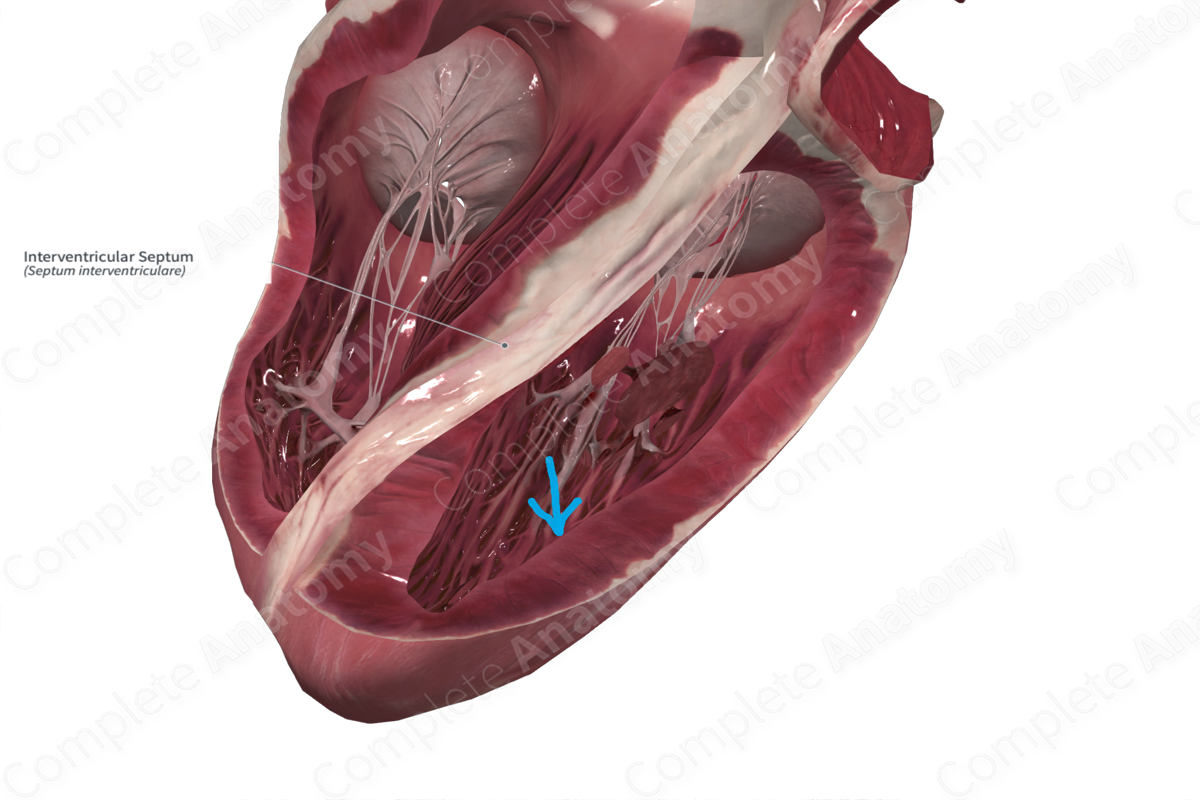
The interventricular septum is a muscular wall that separates the right and left ventricles. The atrioventricular nerve bundle runs through the interventricular septum. The bundle separates into the right and left branches, which run on the right and left sides of the septum respectively, descend towards the apex of the heart as the Purkinje fibers (or subendocardial branches).
Key Featrues/Anatomical Relations
The interventricular septum has a muscular and a membranous portion. The muscular portion forms the majority of the septum and the membranous part is the upper and thinner portion of the septum.
Function
The interventricular septum separates the pulmonary and systemic circulatory systems by ensuring the division of the right and left ventricles. It provides structural support to the heart and contains conducting nerve fibers which ensure coordinated muscular contraction during the cardiac cycle.
List of Clinical Correlates
- Ventricular septal defects
- Tetralogy of Fallot
Learn more about this topic from other Elsevier products
Interventricular Septum

Truncus arteriosus is characterized by a single arterial vessel that originates from the heart, overrides the ventricular septum, and supplies the systemic, coronary, and pulmonary circulations, all from the proximal ascending vessel.
Interventricular Septum: What Is It, Location, and More

The interventricular septum, also known as the ventricular septum, refers to the triangular wall of cardiac tissue that separates the left Learn with Osmosis


