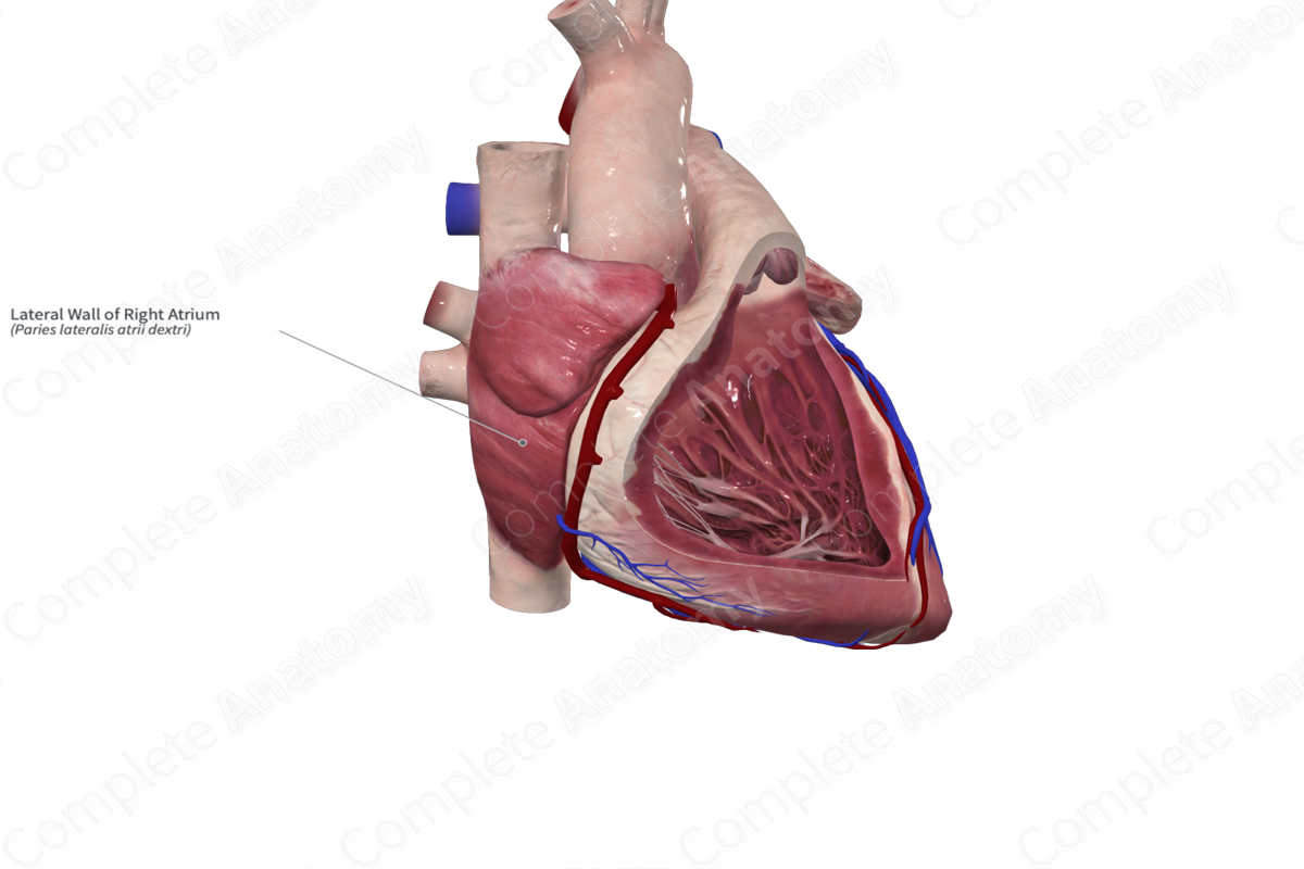
Description
The heart wall windows follow the standard cuts that are made in anatomy dissections to visualize the internal structures and features of the atria and ventricles.
The lateral wall of the right atrium illustrates a horizontal cut that runs along the edge of the right auricle to the superior vena cava. It descends vertically to the inferior vena cava. The cut is then extended along the coronary sulcus.
Once these cuts have been made, the wall of the right atrium is reflected to expose the inner surface of the anterolateral wall, which contains a roughened anterior wall with muscular ridges called the pectinate muscles. There are several other gross anatomical landmarks within the right atrium including:
- the openings of the superior and inferior venae cavae with its valve;
- opening of the valve of the coronary sinus;
- fossa ovalis;
- limbus of fossa ovalis;
- interatrial septum;
- superior aspect of the right atrioventricular valve (or tricuspid valve).
Learn more about this topic from other Elsevier products
Right Atrium

The right atrium (RA) is identified as the chamber that receives the insertion of the inferior vena cava and coronary sinus and by a broad-based triangular appendage with characteristic pectinate muscle morphology extending to the atrioventricular (AV) junction.


