Morphology/Structure
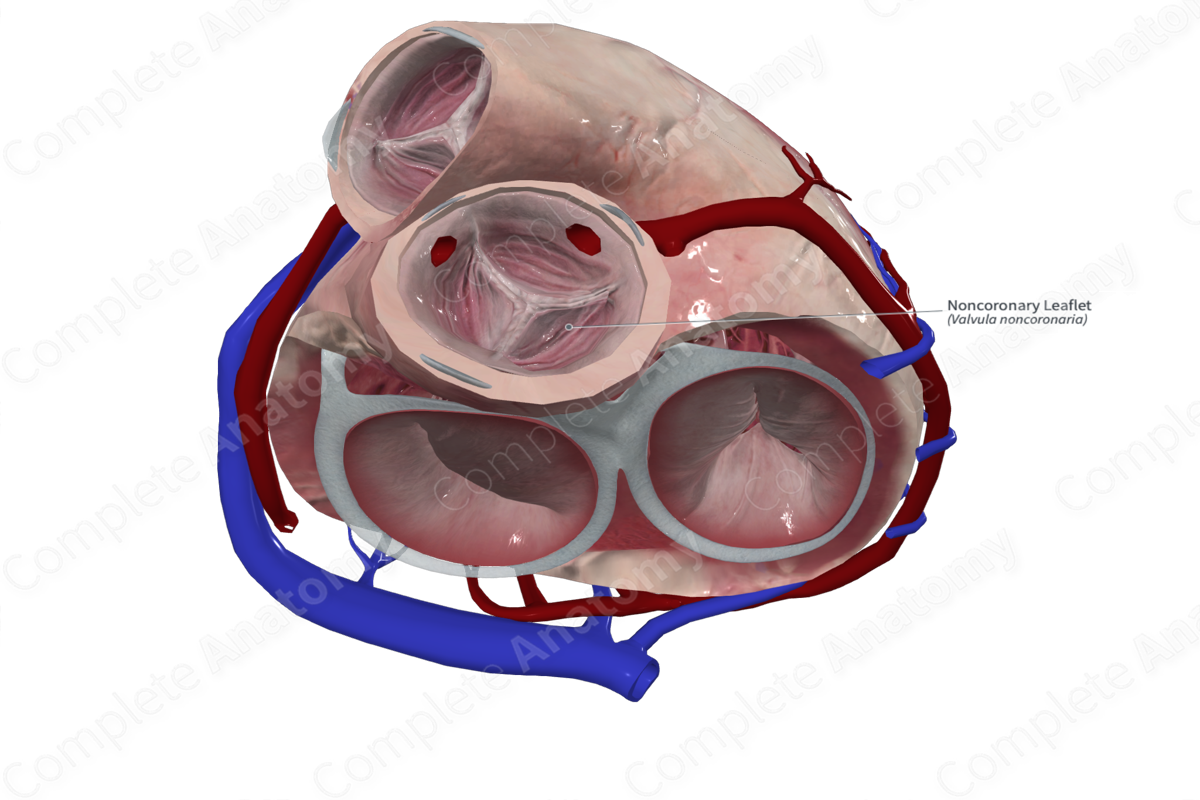
The aortic valve is composed of an anulus and three semilunar leaflets (cusps); the right, left, and noncoronary (nonadjacent) semilunar leaflets.
The leaflets are covered with endocardium and are attached to the wall of the aortic sinuses and supporting structures of the ventricles. The semilunar valves do not require chordae tendineae or papillary muscles to function as their semilunar attachment to the aortic wall and their shape gives them their inherent stability.


Related parts of the anatomy
Key Features/Anatomical Relations
The noncoronary (nonadjacent) leaflet does not have an opening to the coronary vessels. The left semilunar leaflet lies next to the ostia of the left coronary artery and is therefore also known as the left coronary leaflet. The right semilunar leaflet lies adjacent to the ostia of the right coronary artery and is therefore also known as the right coronary leaflet.
Function
The noncoronary (nonadjacent) semilunar leaflet is one of three leaflets forming the aortic valve which permits unidirectional flow of oxygenated blood from the left ventricle to the aorta and prevents retrograde flow.
