Morphology/Structure
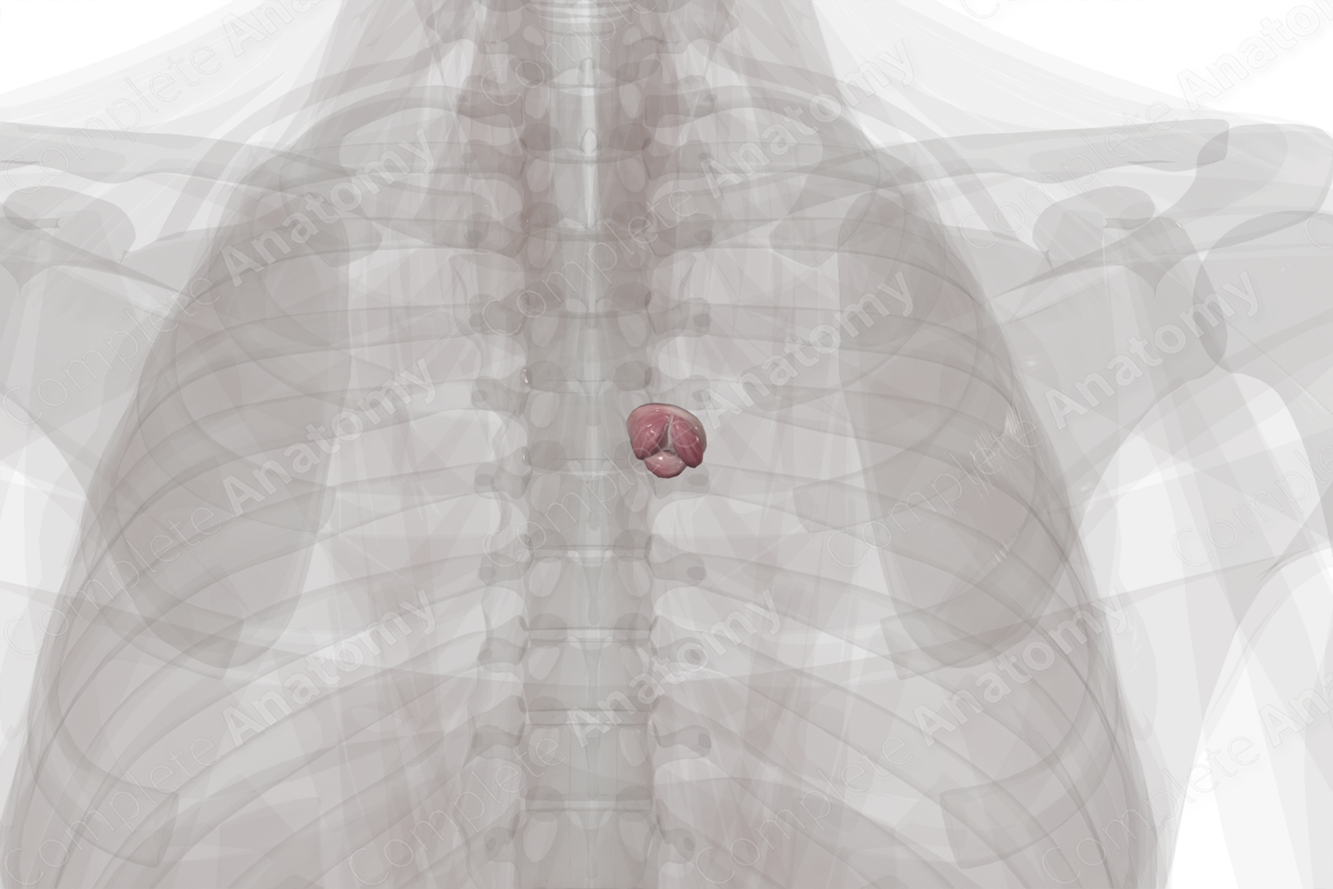
The pulmonary valve (or right semilunar valve) is located at the base of the pulmonary trunk, just above the infundibulum. The pulmonary valve is composed of a fibrous ring with three semilunar leaflets (or cusps); the right, left, and anterior (nonadjacent) semilunar leaflets.
These leaflets are covered with endocardium and are attached to the wall of the infundibulum and supporting structures of the ventricles. The semilunar shape of the leaflets ensures that they’re in close proximity to each other during diastole. To aid in apposition, the free edges of the leaflets are thickened in a region called the lunule, the apex of which is called the nodule.


Key Features/Anatomical Relations
The leaflets are supported within the sinuses and extend from their proximal attachment beyond the free margin of the semilunar leaflets to the sinutubular junction. This is the junction between the pulmonary sinus and the pulmonary trunk. The location of the fibrous rings is often described as the origin of the sinuses, where the hinges of the leaflets attach.
The semilunar valves do not require chordae tendineae or papillary muscles to function, as their semilunar attachment to the wall of the pulmonary trunk gives them their inherent stability. When the valve is closed, the commissures are the points of apposition between adjacent leaflets. Close apposition is essential to the determination of valvular competence.
The pulmonary valve can be auscultated in the second intercostal space, at the left sternal edge below the manubriosternal junction.
Function
The pulmonary valve separates the right ventricle from the pulmonary trunk and permits unidirectional flow of deoxygenated blood from the ventricle to the artery. When the heart is filling during diastole, the leaflets are opposed. Atrial systole may cause a slight movement to the leaflets; however, during ventricular systole, the valves are passively open due to the force of blood leaving the ventricles. The valves close immediately at the end of systole.
List of Clinical Correlates
- Pulmonary valve stenosis
- Pulmonary valve regurgitation
- Infective endocarditis
- Rheumatic heart disease
Learn more about this topic from other Elsevier products
Pulmonary Valve

The pulmonary valve is a semilunar valve situated between the RV and pulmonary artery, has three leaflets, and arises from the infundibulum of the RV.
