Septal Leaflet of Right Atrioventricular Valve
Cuspis septalis valvae atrioventricularis dextrae
Read moreMorphology/Structure
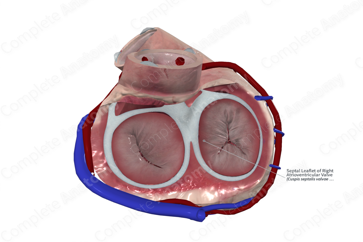
The septal leaflet is one of the three leaflets of the right atrioventricular valve. The leaflets are named according to the regions of the orifice that they attach to and are called the superior, inferior, and septal leaflets. The leaflets have a fibrous core and are coated with endocardium. Each leaflet has a rough, clear, and basal zone. The rough zone corresponds to the region of the free margin where it provides an attachment for the chordae tendineae. It’s this region that comes into contact with the free margins of the adjacent leaflets when the valve is closed. The clear zone is smooth and more opaque and is the site of attachment of occasional chordae tendineae. The basal zone is thicker, due to increased collagen. It’s the region where the leaflets insert into the anulus.


Key Features/Anatomical Relations
The leaflets of the right atrioventricular valve are supported by and attached to chordae tendineae, which in turn attaches to the papillary muscles of the ventricular wall. The septal leaflet is connected to the anterior and septal papillary muscles at the anteroseptal commissure via chordae tendineae.
The attachment of the septal leaflet forms one of the borders of the triangle of Koch, which aids surgeons in the location of the atrioventricular node.
Function
The septal leaflet of the right atrioventricular valve works the superior and inferior leaflets to prevent retrograde blood flow into the right atrium during ventricular systole.
Learn more about this topic from other Elsevier products

