Morphology/Structure
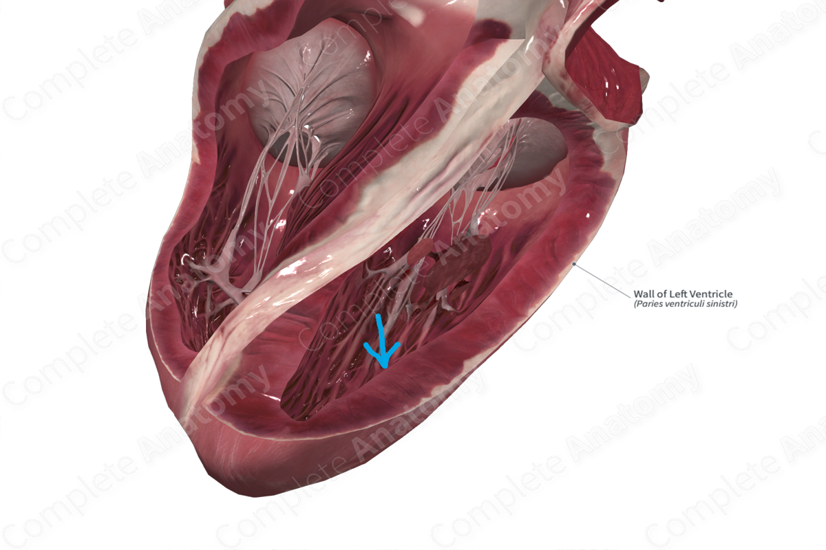
The left ventricle is the dominant chamber in the heart. The wall consists of an inner endocardial layer, a thick myocardial layer (up to 12 mm), and a thin outer epicardial layer. It forms the apex of the heart, and the inferior and a portion of the anterior (sternocostal) surface of the heart.


Key Features/Anatomical Relations
The majority of the interior surface of the left ventricle is covered in irregular columnar projections known as trabeculae carneae.
Emerging from the inner wall are two sets of papillary muscles, the inferoseptal and superoposterior papillary muscles. They’re varied in dimensions and may be bifid. The inferoseptal papillary muscle arises from the inferior (posterior) ventricular wall. The superoposterior papillary muscle arises from the anterior ventricular wall.
The atrioventricular orifice, where the left atrioventricular valve sits as well as the outflow tract leading to the aortic valve both have smooth internal walls.
Function
The left ventricle, with its thick myocardial layer, contracts during ventricular systole, forcing the oxygenated blood through the aortic valve and into the aorta for transport to systemic tissues.
