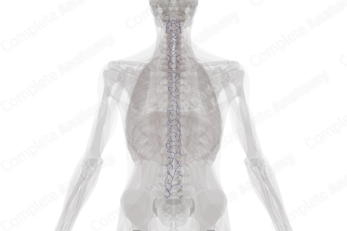
Anterior External Vertebral Venous Plexus
Plexus venosus vertebralis externus anterior
Read moreQuick Facts
Origin: Anterior to the vertebral bodies.
Course: Extends the entire length of the vertebral column.
Tributaries: The basivertebral vein.
Drainage: Vertebrae and local tissue.


Origin
The anterior external vertebral venous plexus is formed by a network of interconnected vessels located anterior to the vertebral bodies.
Course
The anterior external vertebral venous plexus is found along the entire length of the vertebral column, from the foramen magnum to the sacral hiatus. It is most developed in the cervical region.
Tributaries
The anterior external vertebral venous plexus is a valveless network, which is free to anastomose with other components of the vertebral venous plexus. It communicates with the basivertebral and intervertebral veins and venules from local tissues.
Structures Drained
The anterior external vertebral venous plexus drains blood from the vertebrae, as well as muscle and connective tissues located close by.
List of Clinical Correlates
- Arteriovenous malformation
Learn more about this topic from other Elsevier products
Plexus

Visceral plexuses are a network of nerve fiber and ganglia surrounding organs of the abdomen and pelvis region that convey sympathetic, parasympathetic, and visceral afferent input.
