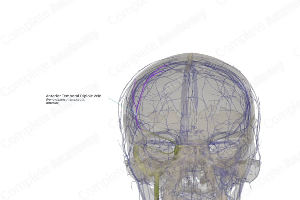
Anterior Temporal Diploic Vein (Left)
Vena diploica temporalis anterior
Read moreQuick Facts
Origin: Confined chiefly to the frontal bone and anterior parietal bone.
Course: Run through the diploic channels between the outer and inner tables of the cortical bone of the parietal and frontal bones near the pterion.
Tributaries: None.
Drainage: Parietal and frontal bone.
Origin
The anterior temporal diploic veins are confined chiefly to the frontal and anterior parietal bones of the skull and are topographically located in an area delimited by the bregma, frontozygomatic suture, pterion, and asterion. There are certain areas in the anterior skull region, where the frontal and anterior temporal diploic veins overlap.
Course
Diploic channels are situated in the diploe or the spongy layer, between the outer and inner tables of the cortical bones. The anterior temporal diploic veins represent a network of valveless intraosseous veins which run dorsocaudally through the diploic channels near the pterion. The pterion is surrounded by multiple small venous channels and the anterior temporal diploic veins have draining points in all pterional areas. The main trunk can be followed upward almost parallel to the border of the squamous temporal bone and coronal sutures. The main trunk of the anterior temporal diploic vein can be seen running towards the midline where it drains into the superior sagittal sinus. It may also pierce the greater wing of sphenoid bone to drain into the sphenoparietal sinus (Garcia-Gonzalez et al., 2009).
Tributaries
There are no named tributaries; however, it does form an anastomosis with the frontal and posterior temporal diploic veins.
Structures Drained
The anterior temporal diploic veins drain the parietal and frontal bones.
References
Garcia-Gonzalez, U., Cavalcanti, D. D., Agrawal, A., Gonzalez, L. F., Wallace, R. C., Spetzler, R. F. and Preul, M. C. (2009) 'The diploic venous system: surgical anatomy and neurosurgical implications', Neurosurg Focus, 27(5), pp. E2.

