Quick Facts
Origin: Continuation of the cranial part of anterior internal vertebral venous plexus or from a primary head vein.
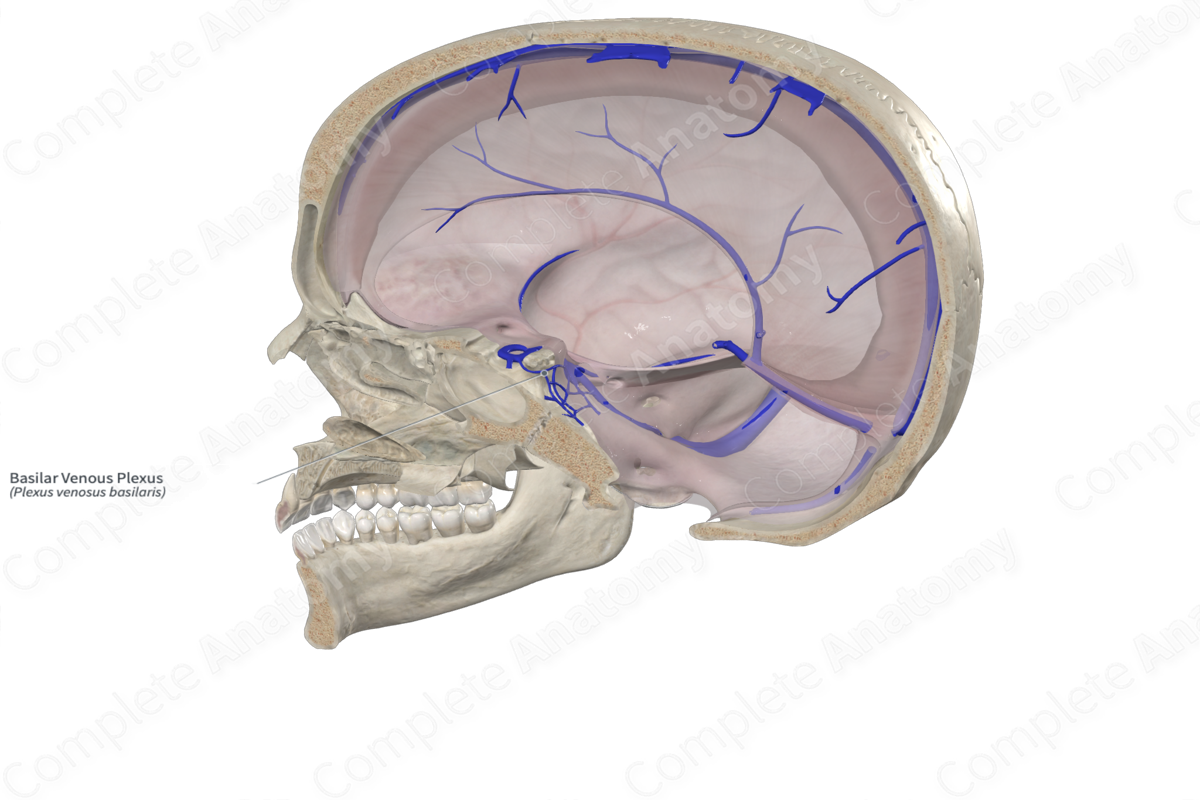
Course: Consists of interconnecting venous channels between layers of dura mater on the clivus.
Tributaries: Numerous connections exist with surrounding venous channels, such as inferior petrosal sinuses, internal vertebral venous plexus, cavernous and superior petrosal sinuses, marginal sinus, venous network of the hypoglossal canal, condylar emissary veins, and the inferior surface of clivus.


Origin
There is contradictory evidence in the literature regarding the origin of the basilar venous plexus. Some believe that it arises as a continuation of the cranial part of the anterior internal vertebral venous plexus. Others found no anatomical link between the two and postulated that the basilar plexus of veins is likely derived from a primary head vein (de Caro et al., 1990).
Course
The basilar sinus and plexus consist of interconnecting venous channels between layers of dura mater on the clivus.
Tributaries
The basilar venous plexus has numerous connections with the venous channels around it. For instance, it interconnects the inferior petrosal sinuses and joins posteroinferiorly with the internal vertebral venous plexus. Anteriorly, it has additional connections with the cavernous and superior petrosal sinuses. If the marginal sinus enlarges, it may also join anteriorly with the basilar plexus of veins. The basilar plexus indirectly communicates with the pterygoid plexus and facial veins through the cavernous sinus and superior ophthalmic vein, respectively.
There are additional intraosseous connections between the basilar plexus and the inferior side of the clivus via the basilar canal and connections with the venous network of the hypoglossal canal and condylar emissary veins (Tubbs et al., 2007).
References
de Caro, R., Parenti, A., Capitanio, G., Ori, C., Bracco, F. and Ricchieri, G. L. (1990) 'Basilar arterio-venous pseudoparallelism due to persistence of embryonal venous pattern', Acta Neurochir (Wien), 104(1-2), pp. 73-6.
Tubbs, R. S., Hansasuta, A., Loukas, M., Louis Jr., R. G., Shoja, M. M., Salter, E. G. and Oakes, W. J. (2007) 'The basilar venous plexus', Clinical Anatomy, 20(7), pp. 755-759.
Learn more about this topic from other Elsevier products

