
Quick Facts
Origin: Union of internal and external iliac veins.
Course: Ascends obliquely to continue as the inferior vena cava at the fifth lumbar vertebra.
Tributaries: Median sacral, ascending lumbar, iliolumbar, internal iliac, and external iliac veins.
Drainage: Pelvic walls, pelvic viscera, and lower limb.


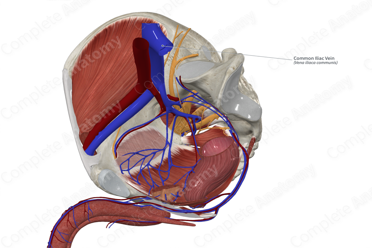

Origin
The common iliac veins are formed from the union of the internal and external iliac vein, anterior to the sacroiliac joint.
Course
The right common iliac vein is much shorter and more vertical than the left common iliac vein. It ascends posteriorly then laterally to the common iliac artery. The left common iliac vein ascends medially then posteriorly to the artery.
Tributaries
Both the left and right common iliac veins receive the iliolumbar and ascending lumbar veins. The median sacral vein drains into the left common iliac vein.
Structures Drained
The common iliac veins drain blood from the pelvic viscera, pelvic walls, and lower limbs.
Learn more about this topic from other Elsevier products
Common Iliac Vein

The common iliac veins are formed by the union of the external and internal iliac veins opposite the sacroiliac joint.
