Quick Facts
Origin: Posterior vertebral venous plexus.
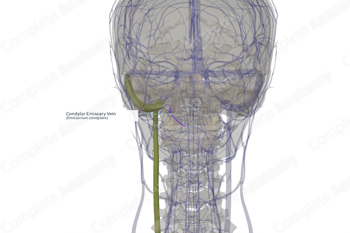
Course: Passes through the condylar fossa of the occipital bone.
Tributaries: None.
Drainage: Suboccipital region.


Related parts of the anatomy
Origin
The posterior condylar emissary vein usually arises from the posterior vertebral venous plexus (part of the suboccipital venous plexus in the posterior neck region).
Course
The posterior condylar emissary vein passes through the posterior condylar fossa to enter the cranial cavity. This condylar fossa is the largest emissary foramen in the retromastoid region of the base of the skull. Inside the cranial cavity, the emissary vein drains into the sigmoid sinus. It could also invariably drain into the marginal or occipital sinuses.
Tributaries
There are usually no tributaries; however, it has been reported that the posterior condylar emissary vein joins the hypoglossal plexus (3.84%) or with internal jugular vein (3.84%) below the jugular foramen (Tubbs, Shoja and Loukas, 2016).
Structures Drained
Venous blood from the posterior scalp region is drained via the posterior condylar emissary vein and is delivered to the sigmoid sinus. However, one must be aware of the fact that emissary veins are valveless channels and hence, the blood can flow in either direction.
References
Tubbs, R. S., Shoja, M. M. and Loukas, M. (2016) Bergman's Comprehensive Encyclopedia of Human Anatomic Variation. Wiley.
Learn more about this topic from other Elsevier products
Vein

A venous sinus is a vein with a thin wall of endothelium that is devoid of smooth muscle to regulate its diameter.
