
Quick Facts
Origin: Within the main portal fissure.
Course: Joins the left hepatic vein and terminates in the inferior vena cava.
Tributaries: Small branches draining segments IV, V and VIII of the liver.
Drainage: Central section of the liver.

Origin
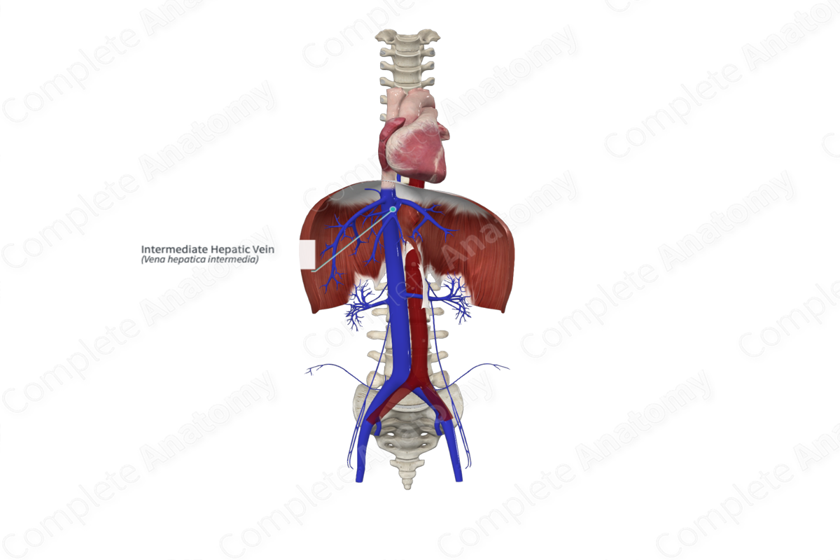
The intermediate (middle) hepatic vein originates in the main portal fissure between the right and left halves of the liver.
Course
The intermediate hepatic vein typically joins the left hepatic vein as it travels posterosuperior through the liver, before it ultimately drains directly into the inferior vena cava (Standring, 2016).
Tributaries
The intermediate hepatic vein receives small branches draining venous blood directly from segments, IV, V, and VIII of the liver.
Structures Drained
The intermediate hepatic vein drains the central aspect of the liver, associated with segments IV, V, and VIII.
List of Clinical Correlates
- Liver transplantation and hepatic resections
References
Standring, S. (2016) Gray's Anatomy: The Anatomical Basis of Clinical Practice. Gray's Anatomy Series 41 edn.: Elsevier Limited.
Learn more about this topic from other Elsevier products
Hepatic Vein

The hepatic vein is composed of three major tributaries (right, middle, and left intrahepatic branches) and drains blood flow from the right, left, and quadrate lobes into the inferior vena cava.
