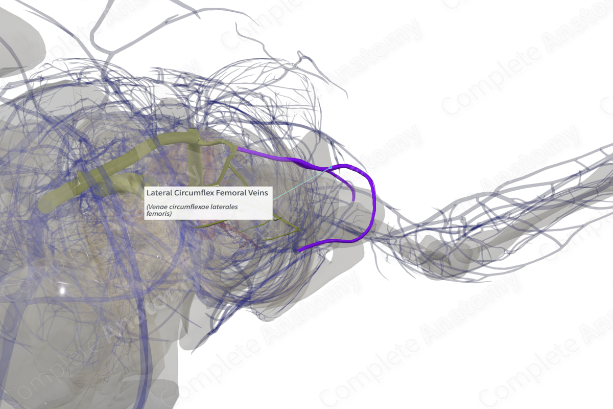
Lateral Circumflex Femoral Veins (Right)
Venae circumflexae laterales femoris
Read moreQuick Facts
Origin: Around the proximal femur.
Course: Horizontal course to drain into the femoral or deep femoral vein.
Tributaries: Retinacular veins.
Drainage: Hip joint and thigh muscles.


Related parts of the anatomy
Origin
The lateral circumflex femoral vein is formed by the union of unnamed ascending, transverse, and descending tributaries, that follow a similar pattern to the branches of the lateral circumflex femoral artery.
Course
From its origin, the lateral circumflex femoral vein takes a horizontal course and runs medially around the proximal shaft of the femur bone to drain into the femoral or deep femoral vein.
Tributaries
The lateral circumflex femoral vein receives the retinacular veins which drain the head and neck of the femur. Additionally, the lateral circumflex femoral vein receives ascending and transverse tributaries. However due to variation they are not officially named. Additionally, it forms an anastomosis with the superficial tributary of medial circumflex femoral vein.
The ascending tributary of the lateral circumflex femoral vein courses inferiorly along the intertrochanteric line at the anterior aspect of the neck of the femur, until it drains into the lateral circumflex vein.
The transverse tributary is the smallest tributary of the lateral circumflex femoral vein. It pierces vastus lateralis muscle and runs anterior to vastus intermedius muscle. It forms anastomoses with the transverse tributary of medial circumflex vein (deep to gluteus maximus muscles), the inferior gluteal vein, and first perforating vein in the thigh.
Structures Drained
The lateral circumflex femoral vein drains the proximal deep thigh, including the head, neck, and proximal shaft of the femur.
Learn more about this topic from other Elsevier products

