Quick Facts
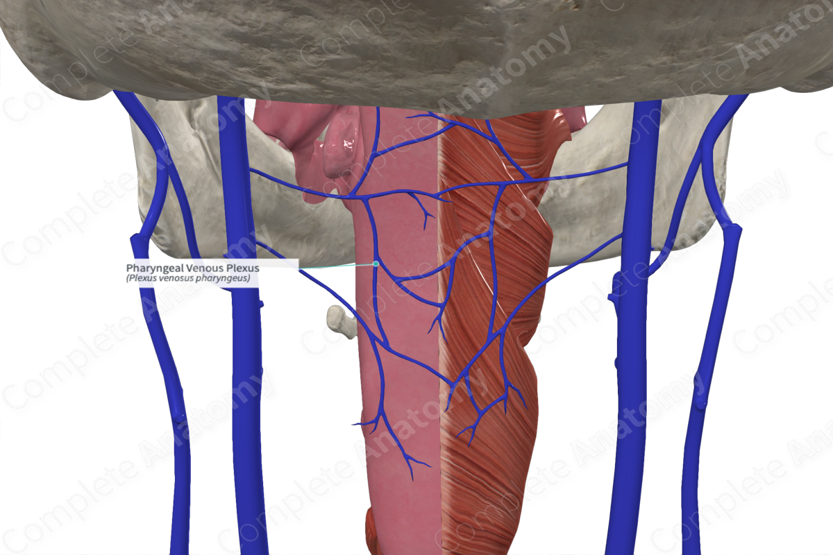
Origin: Union of pharyngeal veins posterolateral to the pharynx.
Course: Passes laterally to the internal jugular vein.
Tributaries: None.
Drainage: Pharynx.


Origin
The pharyngeal plexus is formed by the union of several pharyngeal veins along the posterolateral aspect on the external surface of the pharynx.
Course
The pharyngeal plexus extends laterally to drain into the internal jugular vein. Sometimes, it may drain into the facial, lingual, or superior thyroid veins.
Tributaries
There are no named tributaries; however, the pharyngeal plexus may communicate with the pterygoid plexus. Additionally, emissary veins may connect the pharyngeal plexus with the cavernous sinus, thus connecting intracranial venous sinuses with the extracranial veins (Standring, 2016).
Structures Drained
The pharyngeal plexus drains the pharynx.
References
Standring, S. (2016) Gray's Anatomy: The Anatomical Basis of Clinical Practice., 41st edition. Elsevier Limited.
Learn more about this topic from other Elsevier products
Plexus

Visceral plexuses are a network of nerve fiber and ganglia surrounding organs of the abdomen and pelvis region that convey sympathetic, parasympathetic, and visceral afferent input.
