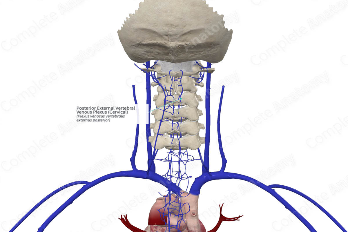
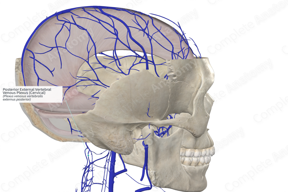
Posterior External Vertebral Venous Plexus (Cervical)
Plexus venosus vertebralis externus posterior
Read moreQuick Facts
Origin: Posterior to the vertebral arches.
Course: Extends the entire length of the vertebral column.
Tributaries: Posterior internal vertebral venous plexus.
Drainage: Vertebrae and local tissue.


Origin
The posterior external vertebral venous plexus is formed by a network of interconnecting vessels running posterior to the vertebral laminae, around and between the spinous process.
Course
The posterior external vertebral venous plexus is a valveless network located along the entire length of the spinal column.
Tributaries
The posterior external vertebral venous plexus communicates with the internal vertebral venous plexus and joins with the vertebral, posterior intercostal, and lumbar veins.
Structures Drained
The posterior external vertebral venous plexus drains the bony tissues and some soft tissues surrounding the spinous processes and laminae.
List of Clinical Correlates
- Arteriovenous malformation
Learn more about this topic from other Elsevier products
Vein

A venous sinus is a vein with a thin wall of endothelium that is devoid of smooth muscle to regulate its diameter.
