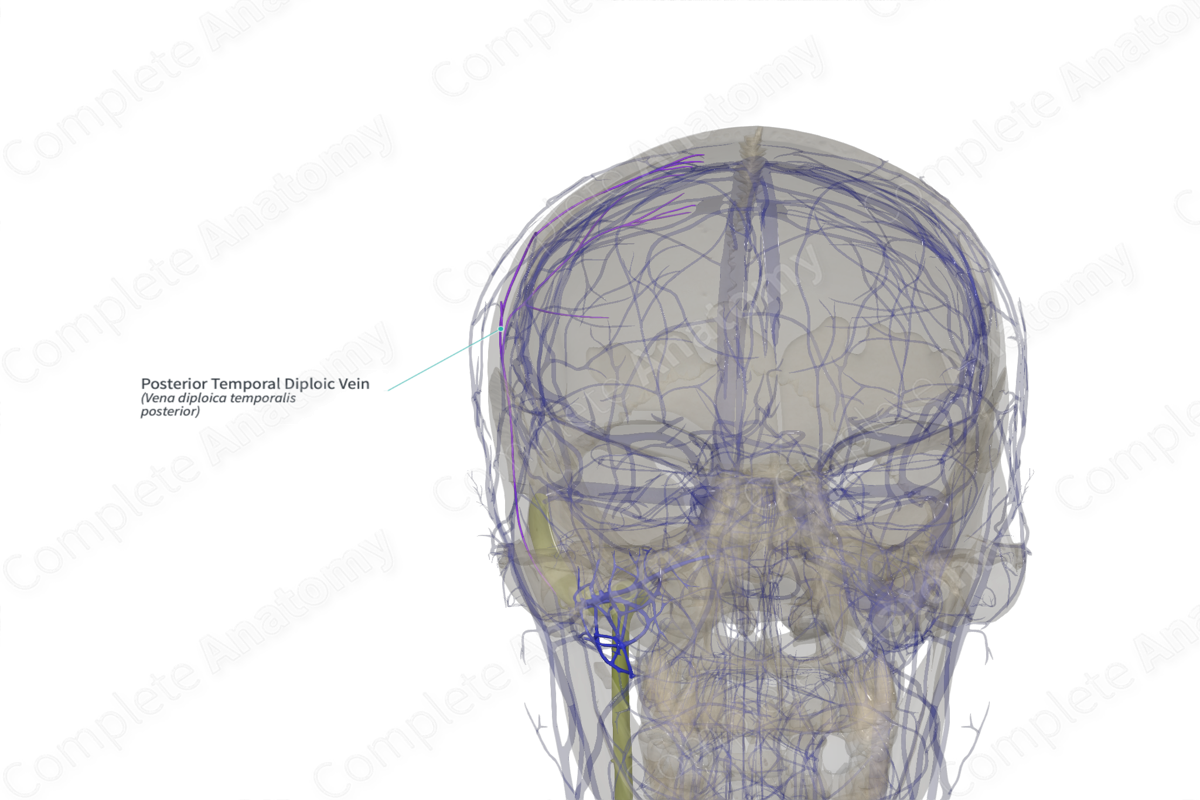
Posterior Temporal Diploic Vein (Right)
Vena diploica temporalis posterior
Read moreQuick Facts
Origin: Situated chiefly in the parietal bone.
Course: Run through the diploic channels between the outer and inner tables of the cortical bone of the parietal bone near the asterion.
Tributaries: None.
Drainage: Parietal bone.

Origin
The posterior temporal diploic vein is located mainly in the parietal bone, in a region which is topographically delimited by the bregma, pterion, asterion, and lambda.
Course
Diploic channels are situated in the diploe or the spongy layer, between the outer and inner tables of the cortical bones. The posterior temporal diploic veins represent a network of valveless intraosseous veins which course primarily through the parietal bone, running anteriorly and dorsomedially relative to the midline. These have a draining point posteriorly close to the asterion or posterosuperior mastoid area (Garcia-Gonzalez et al., 2009).
Tributaries
There are no named tributaries; however, the posterior temporal diploic veins connect with the petrosquamosal sinus, mastoid emissary vein, and sigmoid, transverse, and superior petrosal sinuses to form a complex system that drains the superior sagittal sinus and forms a communication between the extracranial and intracranial venous systems. Sometimes, rich anastomoses exist between the posterior and anterior temporal diploic veins and the occipital diploic veins. Emissary veins, such as the emissary mastoid vein and some parietal and occipital emissary veins, link the posterior temporal and occipital diploic veins with the veins of the posterior scalp and neck (suboccipital venous system).
The posterior temporal diploic veins terminate in the transverse sinus (and sigmoid sinus), through an aperture at the mastoid angle of the parietal bone or through the mastoid foramen (Garcia-Gonzalez et al., 2009).
Structures Drained
The posterior temporal diploic veins drain chiefly the parietal bones of the skull.
References
Garcia-Gonzalez, U., Cavalcanti, D. D., Agrawal, A., Gonzalez, L. F., Wallace, R. C., Spetzler, R. F. and Preul, M. C. (2009) 'The diploic venous system: surgical anatomy and neurosurgical implications', Neurosurg Focus, 27(5), pp. E2.
Learn more about this topic from other Elsevier products

