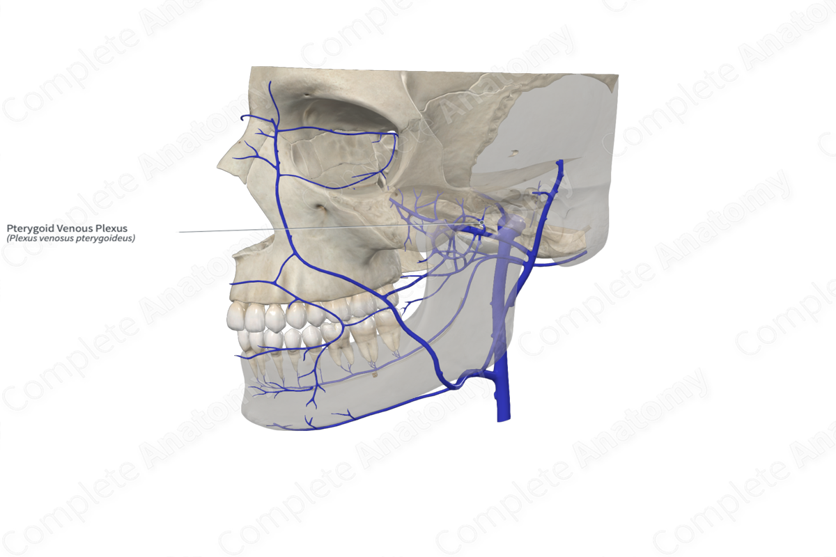
Quick Facts
Origin: Network of veins.
Course: Situated inside the infratemporal fossa, deep to the lateral pterygoid muscle.
Tributaries: Deep temporal, masseteric, buccinator, external palatine, alveolar, parotid, infraorbital, inferior ophthalmic, and sphenopalatine veins, and vein of pterygoid canal.
Drainage: Upper and lower jaws and teeth, muscles of mastication, ear, nose, eye, meninges, and palate.
Related parts of the anatomy
Origin
The pterygoid plexus is a venous plexus formed by a network of veins.
Course
The pterygoid plexus is situated inside the infratemporal fossa, partly deep to the lateral pterygoid muscle.
Tributaries
The pterygoid plexus has several tributaries including sphenopalatine, deep temporal, masseteric, buccinator, external palatine, alveolar, parotid, infraorbital, and inferior ophthalmic veins, and vein of the pterygoid canal.
Anteriorly, the plexus connects with the facial vein via the deep facial vein. Posteriorly, it connects with the retromandibular vein via the maxillary vein. It connects rostrally with the cavernous sinus through emissary veins that pass through the sphenoidal emissary foramen, foramen ovale, and foramen lacerum in the base of the skull to enter the cranial cavity.
Structures Drained
The tributaries of the pterygoid plexus provide venous drainage to the structures which are supplied by the branches of the maxillary artery. Hence, tributaries of the pterygoid plexus provide venous drainage of the nasal cavity, the paranasal sinuses, the nasopharynx, the lateral wall and the roof of the oral cavity, the maxillary and mandibular teeth, and the musculature of the infratemporal fossa. The inferior ophthalmic vein, which drains structures of the bony orbit, has variable anatomy, and may be found as a tributary of the pterygoid plexus.


