
Unfortunately we don't fully support your browser. If you have the option to, please upgrade to a newer version or use Mozilla Firefox, Microsoft Edge, Google Chrome, or Safari 14 or newer. If you are unable to, and need support, please send us your feedback.

Elsevier
Publish with us
Pulmonary Veins of Right Lung
Cardiovascular System

Pulmonary Veins of Right Lung

Venae pulmonalis pulmonis dextri

Read more

Description

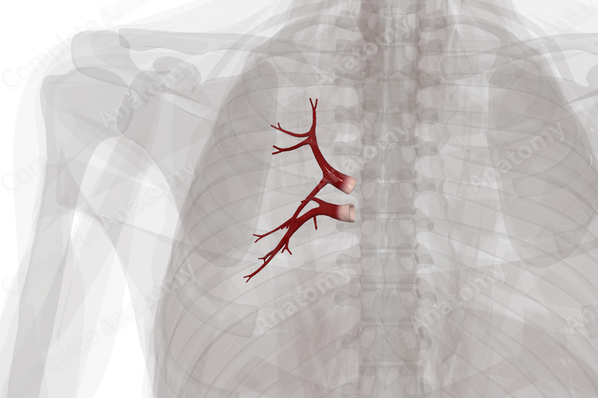
The right lung is drained via the right superior and inferior pulmonary veins. The right superior pulmonary vein is formed by the union of the middle lobe, apical, anterior, and posterior veins, while the right inferior pulmonary vein is formed by the union of superior and common basal veins.

Both the right superior and inferior pulmonary veins travel medially, and pierce through the fibrous pericardium to reach the posterior aspect of the left atrium. The right pulmonary veins are separated from the left counterparts by the oblique pericardial sinus.

Complete Anatomy
The world's most advanced 3D anatomy platform
Try it for Free

List of Clinical Correlates

- Pulmonary vein atresia

- Anomalous pulmonary venous connection

Learn more about this topic from other Elsevier products

Pulmonary Vein

ScienceDirect image

The pulmonary hila, or roots, are complicated structures made up of major bronchi, pulmonary arteries and veins, and hilar lymph nodes.

Explore on ScienceDirectopens in new tab/window

Complete Anatomy

The world's most advanced 3D anatomy platform

Complete Anatomy