Description
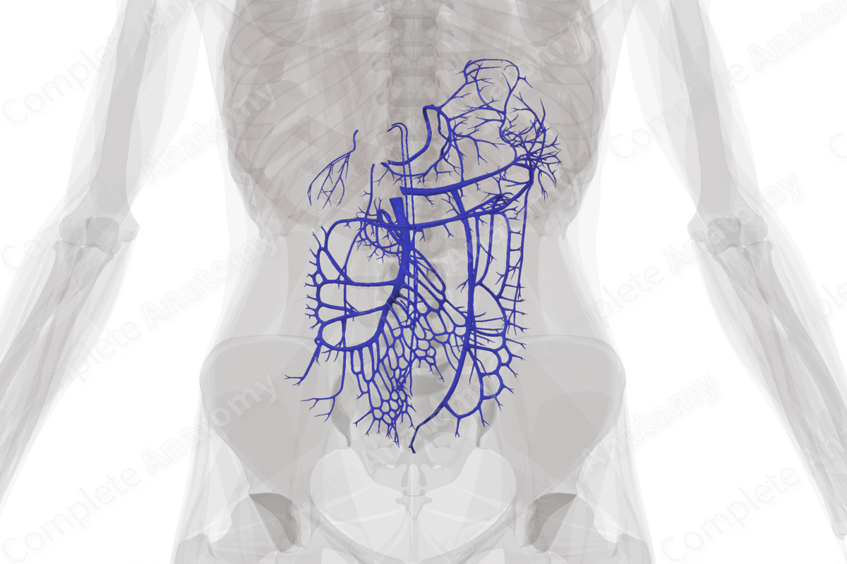
The hepatic portal vein is formed by the union of the superior mesenteric and splenic veins. The latter receives the inferior mesenteric veins. Once formed, the hepatic portal vein has a few direct tributaries including the left and right gastric veins, cystic veins, paraumbilical veins, and the posterior superior pancreaticoduodenal vein.
The portal venous system has tributaries that are at risk of dilating in cases of increased pressure. These include the omental veins, colonic veins, tributaries of the left and right branches of the portal vein, and the superior rectal veins (Standring, 2016).


List of Clinical Correlates
- Cirrhosis
- Portosystemic anastomoses
- Esophageal varices
- Gastric varices
- Rectal varices
- Caput medusae
References
Standring, S. (2016) Gray's Anatomy: The Anatomical Basis of Clinical Practice. Gray's Anatomy Series 41 edn.: Elsevier Limited.
Learn more about this topic from other Elsevier products

