Quick Facts
Origin: Deep palmar arch.
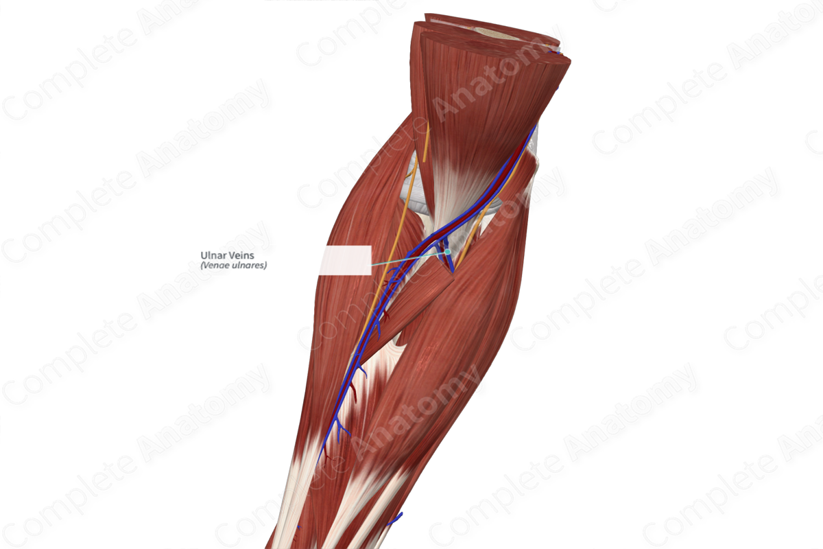
Course: Superiorly, along the medial aspect of the forearm.
Tributaries: Superficial veins of the forearm, anterior and posterior interosseous veins.
Drainage: Hand, medial and posterior aspect of forearm.


Related parts of the anatomy
Origin
The ulnar veins form as the continuation of the medial aspect of the deep palmar arch.
Course
The ulnar veins are the venae comitantes of the ulnar artery and follow the course of the artery along the medial aspect of the forearm. The radial and ulnar veins unite near the elbow and continue as the brachial veins in the arm (Standring, 2016).
Tributaries
There are two sets of venae comitantes in the forearm, the radial and ulnar veins, which receive the superficial and deep palmar venous arches. The deep palmar arch mainly drains into the radial veins. The bulk of the palmar superficial venous blood is shunted to the dorsal aspect of the hand; however, the remainder of blood in the superficial palmar arch primarily drains into the ulnar vein (Doyle and Botte, 2003).
At the wrist, superficial veins connect with the radial and ulnar veins. Along the forearm, the radial and ulnar veins receive tributaries from the anterior and posterior interosseous veins, as well as muscular branches (Standring, 2016, Moore et al., 2013).
Structures Drained
Overall, the ulnar vein and its tributaries contribute to the venous drainage of the forearm via the interosseous veins and through the ulnar vein itself. It also contributes to the drainage of the palmar aspect of the hand.
References
Doyle, J. R. and Botte, M. J. (2003) Surgical Anatomy of the Hand and Upper Extremity. LWW medical book collection: Lippincott Williams & Wilkins.
Standring, S. (2016) Gray's Anatomy: The Anatomical Basis of Clinical Practice. Gray's Anatomy Series 41 edn.: Elsevier Limited.
Learn more about this topic from other Elsevier products

