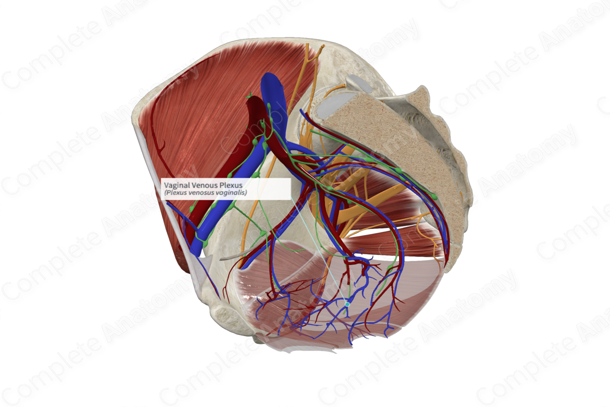
Quick Facts
Origin: Plexus of veins within the vaginal mucosa.
Course: Travel around the sides of the vagina to form anastomoses with the uterine venous plexus.
Tributaries: No tributaries.
Drainage: Vagina.
Related parts of the anatomy
Origin
The vaginal venous plexus is formed as a continuation of the uterine venous plexus.
Course
The vaginal venous plexus surrounds the vaginal walls and unites with the uterine venous plexus (Schünke et al., 2006; Moore, Dalley and Agur, 2013). The uterine venous plexus connects superiorly with the uterine venous plexus and drains into the uterine vein. However, the vaginal venous plexus may drain directly into a left and right vaginal vein (not modeled). If present, the vaginal veins travel superiorly within the pelvis to drain into the uterine vein or take a longer course to drain directly into the internal iliac vein (Standring, 2016).
Tributaries
The vaginal venous plexus does not receive any tributaries, but it does connect superiorly with the uterine venous plexus.
Structures Drained
The vaginal venous plexus drains the walls of the vagina. It may also provide venous drainage to the lower limb (Standring, 2016).
References
Moore, K. L., Dalley, A. F. and Agur, A. M. R. (2013) Clinically Oriented Anatomy. Clinically Oriented Anatomy 7th edn.: Wolters Kluwer Health/Lippincott Williams & Wilkins.
Schünke, M., Schulte, E., Ross, L. M., Lamperti, E. D. and Schumacher, U. (2006) Atlas of Anatomy: Neck and Internal Organs. Thieme Atlas of Anatomy: Thieme.
Standring, S. (2016) Gray's Anatomy: The Anatomical Basis of Clinical Practice. Gray's Anatomy Series 41 edn.: Elsevier Limited.


