Quick Facts
Origin: Union of various venous tributaries from the cochlear part of the inner ear.
Course: Runs parallel to the cochlear aqueduct before draining into the inferior petrosal sinus.
Tributaries: Common modiolar and vestibulocochlear veins.
Drainage: Cochlea and semicircular canals.
Related parts of the anatomy
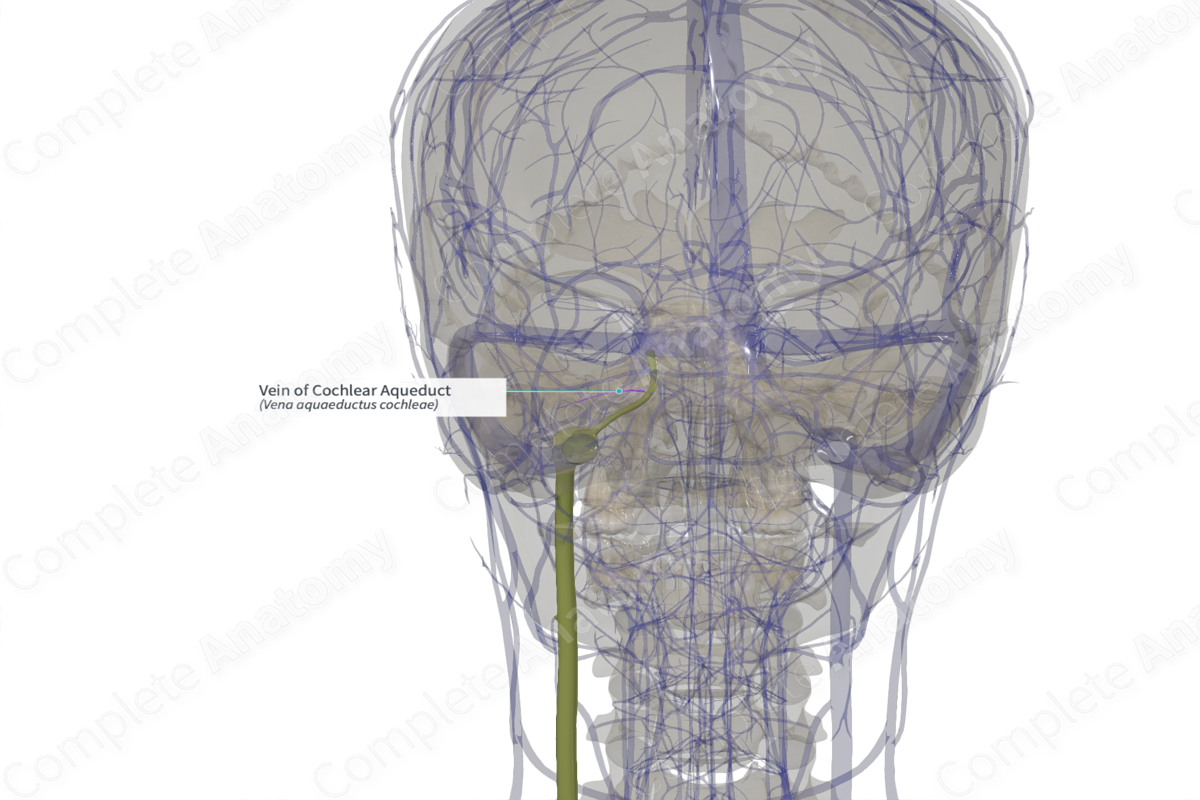
Origin
The vein of the cochlear aqueduct is formed by the union of various venous tributaries from the cochlear part of the inner ear.
Course
The vein of the cochlear aqueduct runs parallel to the cochlear aqueduct by the side of the middle ear, prior to its opening into the inferior petrosal sinus.
Tributaries
The vein of the cochlear aqueduct receives blood from the various venous tributaries, including the common modiolar vestibulocochlear veins. The confluence of these venous tributaries is located at the bottom of the scala tympani near the round window, prior to their opening into the vein of cochlear aqueduct.
The vein of the cochlear aqueduct also receives two communicating veins from the mucoperiosteum of the middle ear. One arises from the mucoperiosteum of the cochlear surface, which runs parallel to the vein of cochlear aqueduct, before draining into it, while the other is from a network of mucoperiosteal vessels near the round window that communicate directly with the vein of cochlear aqueduct (Watanabe, Nakashima and Yanagita, 1988).
Structures Drained
The vein of the cochlear aqueduct receives blood from the scala vestibuli and tympani via the common modiolar veins, and from the cochlea, saccule, and semicircular canals via the vestibulocochlear vein.
References
Watanabe, Y., Nakashima, T. and Yanagita, N. (1988) 'Venous communications of the cochlea after acute occlusion of the vein of the cochlear aqueduct', Arch Otorhinolaryngol, 245(6), pp. 340-3.
Learn more about this topic from other Elsevier products
Vein

A venous sinus is a vein with a thin wall of endothelium that is devoid of smooth muscle to regulate its diameter.
