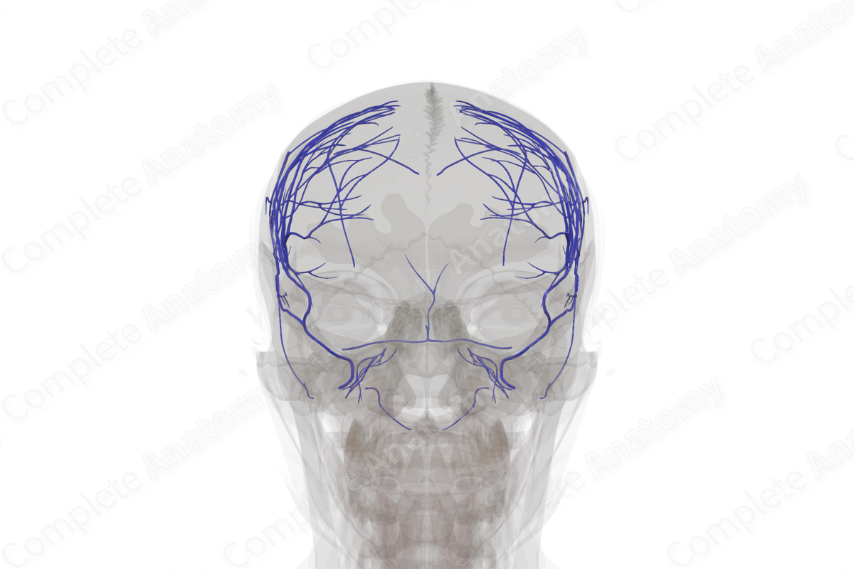
Veins of Meninges & Cranium
Description
The meninges and cranium receive venous drainage from several sources, including the dural venous sinuses, the diploic veins, and the emissary veins.
The diploic veins are a valveless intraosseous network of veins draining the skull itself. These are situated in the diploe or the spongy layer of bone in the cranial vault and run inside the diploic channels, between the inner and the outer layers of the cortical bone of the skull.
The diploic veins communicate with the dural venous sinuses and meningeal veins along the compact inner table of the skull and with the pericranial veins on the outer table via emissary veins (Jivraj et al., 2009; Garcia-Gonzalez et al., 2009).
The emissary veins are small veins which pass through various foramina inside the skull to connect the intracranial venous sinuses with the extracranial veins. Learning their anatomy has clinical implications as these venous channels could provide a route for the spread of infection from outside the cranium into the venous sinuses intracranially, e.g., the spread of infection from the paranasal sinuses to the cavernous sinus.
Related parts of the anatomy
References
Garcia-Gonzalez, U., Cavalcanti, D. D., Agrawal, A., Gonzalez, L. F., Wallace, R. C., Spetzler, R. F. and Preul, M. C. (2009) 'The diploic venous system: surgical anatomy and neurosurgical implications', Neurosurg Focus, 27(5), pp. E2.
Jivraj, K., Bhargava, R., Aronyk, K., Quateen, A. and Walji, A. (2009) 'Diploic venous anatomy studied in-vivo by MRI', Clin Anat, 22(3), pp. 296-301.
Learn more about this topic from other Elsevier products
Vein

A venous sinus is a vein with a thin wall of endothelium that is devoid of smooth muscle to regulate its diameter.


