Description
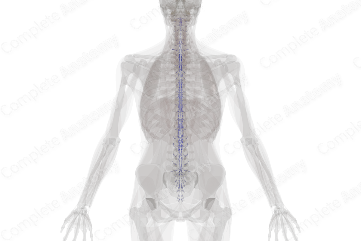
A dense venous plexus (pial or coronal plexus) is located on the surface of the spinal cord. This plexus feeds into the anterior and posterior spinal veins that run longitudinally along the spinal cord. The anterior spinal veins consist of a central vein in the anterior median fissure accompanied by two anterolateral channels found just posterior to the anterior roots of the spinal cord. The posterior spinal veins consist again of a central vein in the posterior median septum and two lateral channels just posterior to the posterior roots of the spinal cord. The spinal veins receive tributaries through the central sulcus of the spinal cord and the pial plexus, thus draining the spinal cord. The spinal veins are drained by the segmental medullary veins into the intervertebral veins, which in turn drain into the vertebral, caval, or azygos systems (Standring, 2016).


Related parts of the anatomy
References
Standring, S. (2016) Gray's Anatomy: The Anatomical Basis of Clinical Practice. Gray's Anatomy Series 41 edn.: Elsevier Limited.
