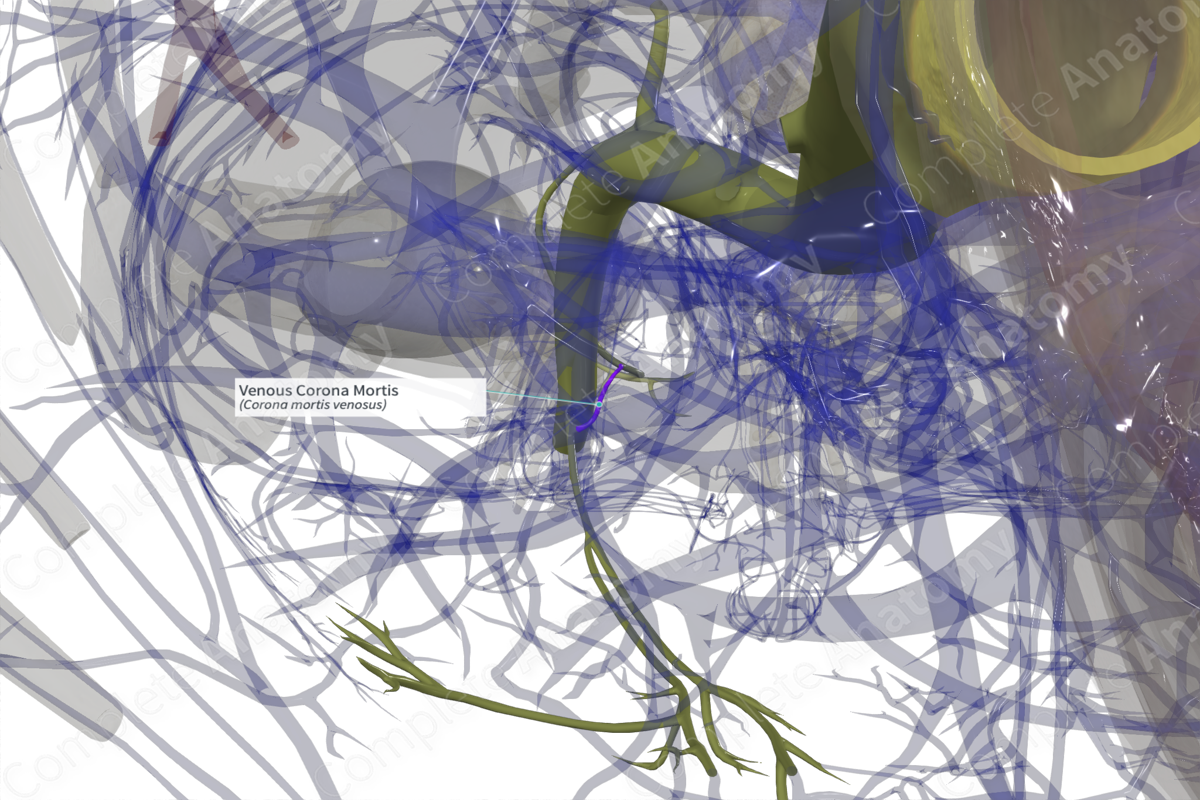
Quick Facts
Origin: Between the obturator vein and the external iliac or inferior epigastric veins.
Course: Passes behind the superior pubic ramus.
Tributaries: None.
Drainage: Anastomosis between two veins.
Origin
The venous corona mortis, or aberrant obturator vein, is an anastomosis formed between the obturator vein and the external iliac or inferior epigastric veins. Although considered an anatomical variant, one study noted its presence in 41.7% of specimens (Sanna et al, 2018).
Course
The corona mortis passes behind the superior pubic ramus, 40-96 mm away from the pubic ramus (Darmanis et al., 2007).
The term “corona mortis” translates as "crown of death," indicating the importance of this structure in orthopedic surgery. Accidental damage to this structure can cause significant hemorrhaging which may be difficult to then achieve hemostasis. It must therefore be considered cautiously during surgery.
Tributaries
There are no named tributaries.
Structures Drained
The corona mortis forms an anastomosis between two major veins draining the pelvis and the anterior abdominal wall.
References
Darmanis, S., Lewis, A., Mansoor, A. & Bircher, M. (2007) Corona mortis: an anatomical study with clinical implications in approaches to the pelvis and acetabulum. Clin Anat, 20(4), 433-9.
Sanna, B., Henry, B. M., Vikse, J., Skinningsrud, B., Pekala, J. R., Walocha, J. A., Cirocchi, R. & Tomaszewski, K. A. (2018) The prevalence and morphology of the corona mortis (Crown of death): A meta-analysis with implications in abdominal wall and pelvic surgery. Injury, 49(2), 302-308.


