Quick Facts
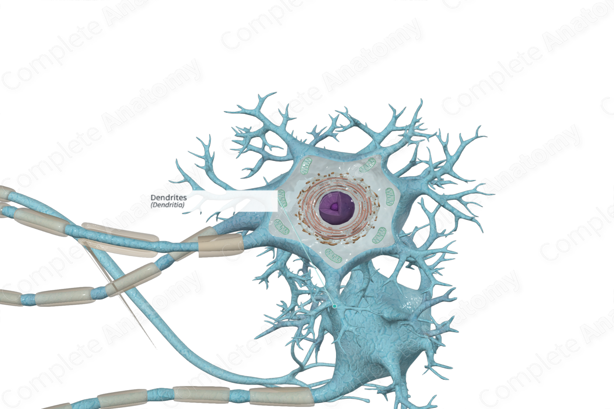
A dendrite is one of the threadlike extensions of the cytoplasm of a neuron, which typically branch into tree-like processes. In unipolar and bipolar neurons, there is a single dendrite, which proximally resembles an axon but branches distally; in multipolar neurons there are many short, branching dendrites. Dendrites form most of the receptive surface of a neuron (Dorland, 2011).
Structure and/or Key Features
Dendrites are short, tree-like, highly branched processes, generally radiating from the cell body, increasing the receptive area of the cell. Dendrites are unmyelinated, usually tapered, and their diameter is greater than the diameter of the neuronal axon. Dendrites become thinner as they branch. The cytoplasm of the dendrites near the cell body contains Nissl granules, ribosomes, rough endoplasmic reticulum, smooth endoplasmic reticulum, mitochondria, microtubules, neurofilaments, and actin filaments. However, they do not contain Golgi complex (Ross and Pawlina, 2006).
Dendrites are overproduced during embryonic development but become reduced in number and size later on (Splittgerber, 2018).
Small mushroom shaped “dendritic spines,” 1–3 μm long and less than 1 μm wide, are connected to the dendrite process.
Anatomical Relations
Dendrites generally radiate from the neuron cell body.
Function
Dendrites function as receptors to enable a single neuron to receive and integrate information arriving on numerous axon terminals from other nerve cells or the external environment. Dendritic spines house the synapses communicating with the neuron from other nerve cell processes.
Clinical Correlates
General anesthetics block synaptic transmission.
References
Dorland, W. (2011) Dorland's Illustrated Medical Dictionary. 32nd edn. Philadelphia, USA: Elsevier Saunders.
Ross, M. H. and Pawlina, W. (2006) Histology: A text and atlas. Lippincott Williams & Wilkins.
Splittgerber, R. (2018) Snell's Clinical Neuroanatomy. Wolters Kluwer Health.
