Quick Facts
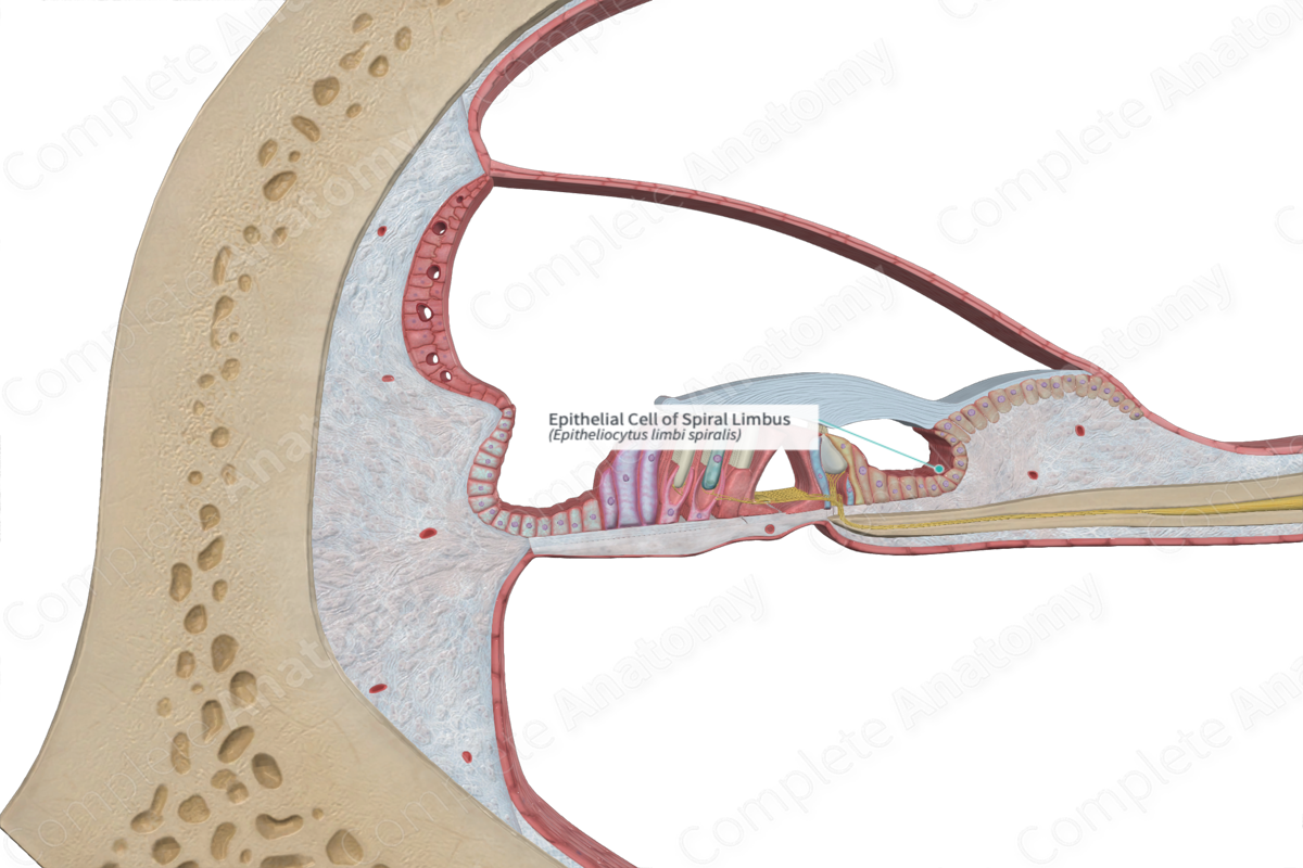
The upper surface of the spiral limbus contains cells that project outwards and are separated at right angles by furrows, resembling teeth, thus are called the acoustic teeth. The spiral limbus is also covered in epithelium, where the cells overlying the acoustic teeth are squamous, while the cells that sit in between, in the interdental furrows, are flask shaped. These are known as interdental cells. This epithelium is continuous with the vestibular membrane and with the epithelium lining the internal spiral sulcus (Standring, 2016).
Structure and/or Key Feature(s)
The upper surface of the spiral limbus contains cells that project outwards and are separated at right angles by furrows, resembling teeth, thus are called the acoustic teeth. The spiral limbus is also covered in epithelium, where the cells overlying the acoustic teeth are squamous, while the cells that sit in between, in the interdental furrows, are flask shaped. These are known as interdental cells. This epithelium is continuous with the vestibular membrane and with the epithelium lining the internal spiral sulcus (Standring, 2016).
The internal spiral sulcus is the C-shaped concavity on the spiral limbus. A stratified squamous epithelium lines the sulcus and continues up over the vestibular edge and across the auditory teeth.
Function
The interdental cells of the spiral limbus produce and maintain tectorial membrane components from a developmental stage. The tectorial membrane is a gelatin sheet which is involved in the auditory signal transduction process via disruption of the stereocilia of the specialized hair cells.
References
Standring, S. (2016) Gray's Anatomy: The Anatomical Basis of Clinical Practice., 41st edition. Elsevier Limited.
