Quick Facts
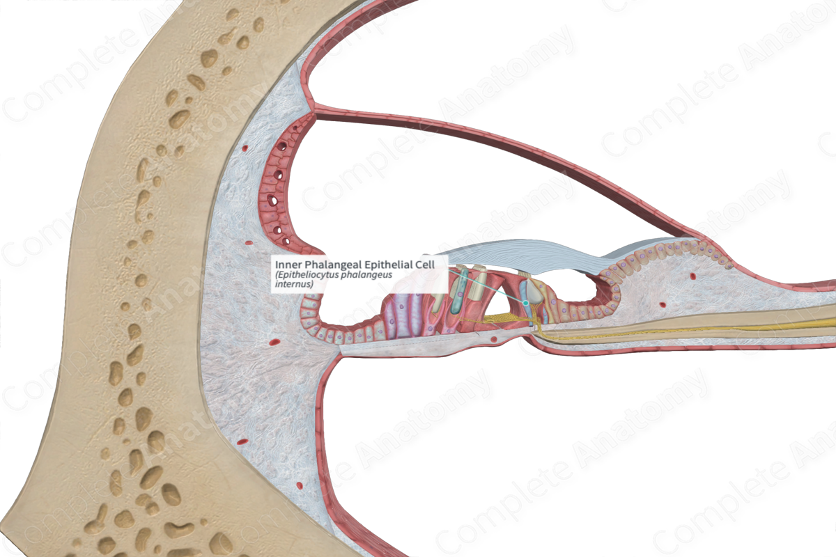
The phalangeal cells are elongated supporting cells of the organ of Corti with bases that rest on the basilar membrane adjacent to the pillar cells. Inner phalangeal cells are arranged in a row on the inner surface of the inner pillar cells and surround the inner hair cells; outer phalangeal cells (or Deiters cells) support the outer hair cells (Dorland, 2011).
Structure and/or Key Feature(s)
The inner phalangeal epithelial cells are one of the main epithelial supporting cells that assist in the positioning of the inner hair cell. The remaining two being the inner border cells and the inner pillar cells. Similar to the other supporting cells, they extend from the basilar membrane to reach the apical aspect of the hair cells, thus spanning the full depth of the sensory epithelium.
The inner phalangeal epithelial cells have a thin, uneven, softly defined rectangular shape. Their apical surface is slanted, continuing on from the slope of the inner border cells. The upper portion of the inner phalangeal epithelial cells contours to accommodate the curved body of the inner hair cell. The apices of the inner phalangeal cells are connected to the neighboring inner hair cells and inner border cells via tight junctions, adherens junctions, and desmosomes. This forms an impermeable barrier, called the reticular lamina, for the passage of ions. Endolymph (high potassium and low sodium concentrations) engulfs the apical surfaces of the hair cells and the supporting cells, while perilymph (high sodium and low potassium) bathes their lateral and basal aspects. This separation generates an electrochemical gradient along the lamina, forming an endocochlear potential which is necessary for the depolarization of hair cells (Standring, 2016).
Anatomical Relations
The inner phalangeal epithelial cells, along with the other supporting cells, are inferior to the under surface of the tectorial membrane, and superior to the basilar membrane. They are interfacing the inner border cells and the important inner hair cells.
Function
The inner phalangeal epithelial cells, along with the other inner supporting cells, contribute to maintenance of the structural organization of the inner hair cells. Additionally, when an inner hair cell is lost due to disease, old age, or excessive noise, the adjacent phalangeal surfaces can expand to cover the gap created, resulting in a characteristic scar.
References
Dorland, W. (2011) Dorland's Illustrated Medical Dictionary. 32nd edn. Philadelphia, USA: Elsevier Saunders.
Standring, S. (2016) Gray's Anatomy: The Anatomical Basis of Clinical Practice., 41st edition. Elsevier Limited.
