Quick Facts
The phalangeal cells are elongated supporting cells of the organ of Corti with bases that rest on the basilar membrane adjacent to the pillar cells. Inner phalangeal cells are arranged in a row on the inner surface of the inner pillar cells and surround the inner hair cells; outer phalangeal cells (or Deiters cells) support the outer hair cells (Dorland, 2011).
Structure and/or Key Feature(s)
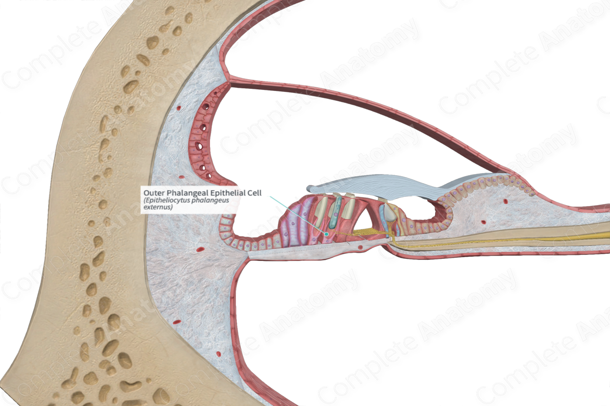
The outer phalangeal epithelial cells are one of the main epithelial supporting cells that assist in the positioning of the outer hair cells. The other supporting cells include the columnar external supporting cells and outer pillar cells. Similar to the other supporting cells, they extend from the basilar membrane to reach the apical aspect of the hair cells, thus spanning the full depth of the sensory epithelium.
The outer phalangeal epithelial cells are irregularly shaped. The basal regions are broad and stretch across the basilar membrane. They possess a sturdy cytoskeleton, composed of clusters of microtubule stalks, granting solid support to the hair cells.
The apical region of each outer phalangeal cell possesses two notable structural aspects. Firstly, the upper aspect of each outer phalangeal epithelial cell is contoured around the base of the rod-like outer hair cell, partially enveloping it in order to support its. In addition, each phalangeal epithelial cell provides long, thin projections which extend obliquely upwards between the individual hair cells. These projections expand out to plug in the spaces between the apices of the hair cells. The apices of the cells are linked by tight junctions, adherens junctions, and desmosomes. This forms an impermeable barrier, called the reticular lamina, for the passage of ions. Endolymph (high potassium and low sodium concentrations) engulfs the apical surfaces of the hair cells and the supporting cells, while perilymph (high sodium and low potassium) bathes their lateral and basal aspects. This separation generates an electrochemical gradient along the lamina, forming an endocochlear potential which is necessary for the depolarization of hair cells (Standring, 2016).
Anatomical Relations
The outer phalangeal epithelial cells, along with the other cells that support the outer hair cells, are inferior to the under surface of the tectorial membrane, and superior to the basilar membrane. They lie in between the rows of outer hair cells, interjacent to the outer pillar cells and the columnar external supporting cells. Some spaces still remain between the outer phalangeal cells and the outer hair cells in which the perilymph fills. This is known as the outer tunnel or the spaces of Nuel.
Function
The outer phalangeal epithelial cells provide a variety of functions, all of which are involved in the structural fixation of the outer hair cells. Along with the outer pillar cells, they form a structural framework which allows for the mechanical stimulation of the hair cells. This is due to the structural makeup of each phalangeal epithelial cell, which is a specialized array of the groups of microtubular stalks, providing a robust support to each hair cell. As the outer hair cells are situated on the concave apical surface of each phalangeal cell, they are effectively rooted onto the basilar membrane via the basal aspect of phalangeal cells. This results in shearing movements between the stereocilia of each hair cell and the undersurface of the tectorial membrane.
The outer phalangeal epithelial cells also fill the vacant areas left by the loss of hair cells. The projections that rise upwards already plug in the spaces between the hair cells and can expand even wider to cover a greater gap in the case of hair cell loss, typically caused by trauma. This helps to restore the function of the reticular lamina but disrupts its typical appearance (Standring, 2016).
References
Dorland, W. (2011) Dorland's Illustrated Medical Dictionary. 32nd edn. Philadelphia, USA: Elsevier Saunders.
Standring, S. (2016) Gray's Anatomy: The Anatomical Basis of Clinical Practice., 41st edition. Elsevier Limited.
