Quick Facts
The pillar epithelial cells are elongated supporting cells found in the organ of Corti in a double row (inner and outer pillar cells), with their heads joined and their bases on the basilar membrane widely separated so as to form a tunnel that extends the length of the cochlea (Dorland, 2011).
Structure and/or Key Feature(s)
The pillar cells are epithelial structures located on the basilar membrane in the cochlear duct. There are two types of pillar cells, the inner pillar cells and the outer pillar cells, and these are anatomically and functionally related to the inner and outer hair cells respectively.
The pillar cells are the most centrally positioned epithelium in the spiral organ of Corti. There are nearly six thousand inner pillar cells and close to four thousand outer pillar cells. The cells are irregularly shaped and are aptly named given that the apical and basal regions are wider than the lengthy narrow body.
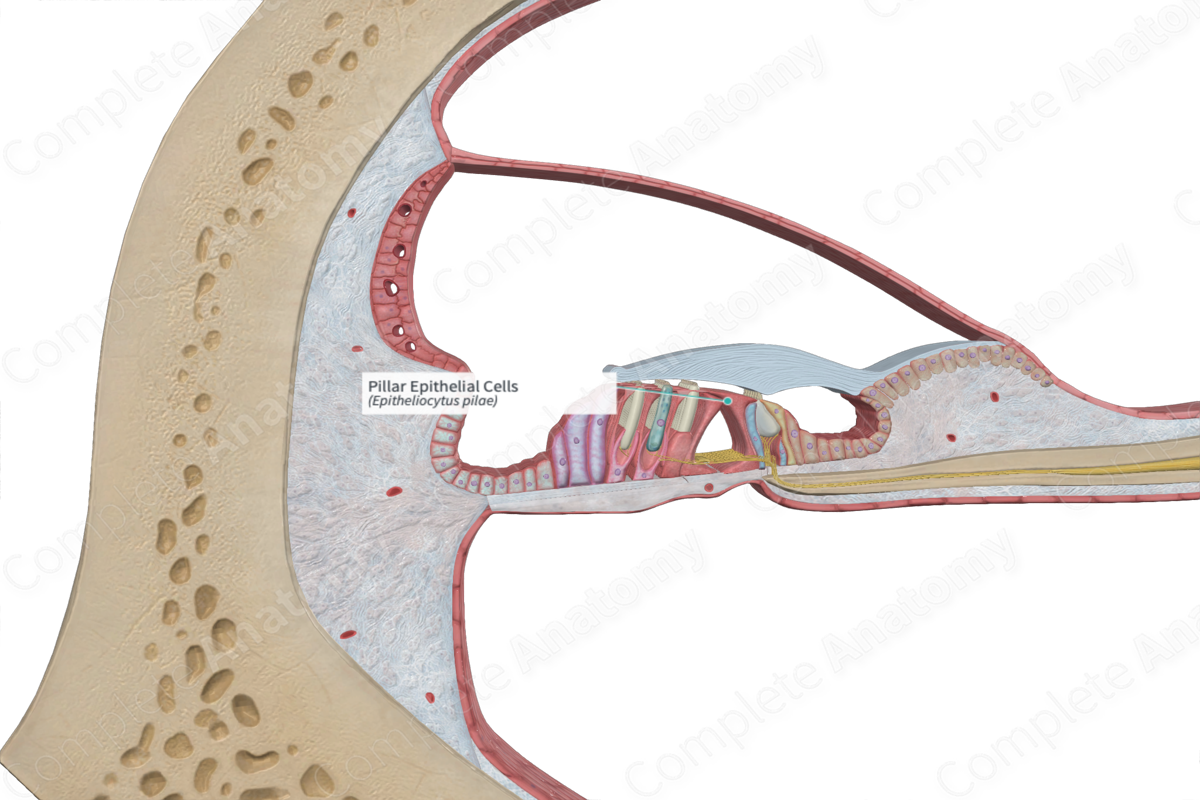
Each pillar cell has a crus (base), long scapus (rod-like body), and caput (head). Each cell is comprised of microtubules which originate in the crus. The microtubules are bundled together in parallel in the scapus. The microtubules eventually branch out and terminate in the caput. The nucleus of each pillar cell is located in the crus (Standring, 2016).
The crus is broad enough so that the crus of the inner and outer pillar cells are in contact with each other. The scapus is thin and rod-like and is well distanced from the other pillar cell bodies. The caput (head) has a similar stretched out appearance as the crus. The caput of the inner pillar cell has a greater width than that of the outer pillar cell and overhangs it. This overhang bears grooves for the insertion of the outer pillar cell caput. The inner and outer pillar cells are inclined toward each other. The scapus of an internal pillar cell inclines at an angle of sixty degrees with the basilar membrane, while those of the outer pillar cells incline at an angle of forty degrees with the basilar membrane.
In short, the broad crus and caput of each inner pillar cell make contact with the opposing outer pillar cell, while the scapus of each cell is separated. This results in the formation of the upper borders of a small triangular shaped channel known as the inner tunnel of Corti. The basilar membrane forms the floor of this tunnel. The outer tunnel is filled with perilymph, also known as corticolymph, which comes from the scala tympani and diffuses through the basilar membrane (Standring, 2016).
Anatomical Relations
The pillar cells are inferior to the under surface of the tectorial membrane and superior to the basilar membrane. The crus of each pillar cell is situated on the surface of the basilar membrane, while the caput is located nearer to the tectorial membrane.
The inner pillar cells are external to the row of inner hair cells and the inner phalangeal epithelial cells surrounding them. The outer pillar cells are external to the inner pillar cells. The caput of the outer pillar cells inserts into the groove on the underside of the caput of the inner pillar cells. The outer pillar cells are internal to the outer hair cells, the outer phalangeal epithelial cells, and the outer supporting cells.
The inner tunnel of Corti is formed by three borders; the bodies of the inner and outer pillar cells and the zona arcuata of the basilar membrane.
Function
The function of the pillar cells is in no doubt due to the unique and robust shape of the cells. Given their proximity and direct contact to the hair cells, they help stabilize the position of these cells, providing a structural frame that allows for stimulation of the hair cells and protects against interruption to the synaptic contacts at the ease of each hair cell. With their strong cytoskeletons and lengthy apical and basal processes, they maintain the structural integrity of the inner tunnel of Corti, even against the added pressure of the surrounding cells during acoustic stimulation events (Wan, Corfas and Stone, 2013).
References
Dorland, W. (2011) Dorland's Illustrated Medical Dictionary. 32nd edn. Philadelphia, USA: Elsevier Saunders.
Standring, S. (2016) Gray's Anatomy: The Anatomical Basis of Clinical Practice., 41st edition. Elsevier Limited.
Wan, G., Corfas, G. & Stone, J. S. (2013) Inner ear supporting cells: rethinking the silent majority. Seminars in cell & developmental biology, 24(5), 448-459.
