Quick Facts
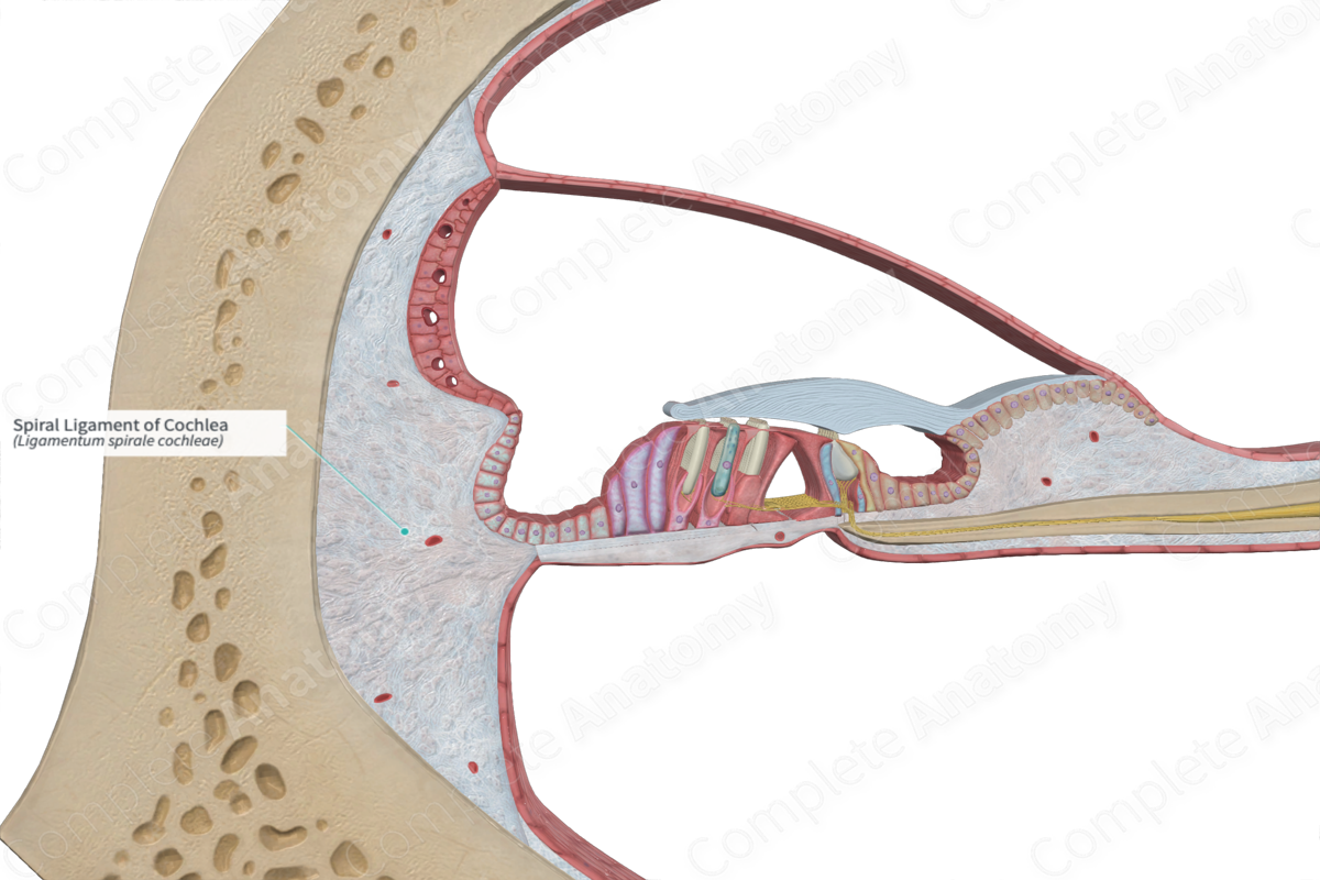
The spiral ligament of cochlea is a band of thickened periosteum in the bony cochlea (Dorland, 2011).
Structure and/or Key Feature(s)
The spiral ligament is a large fibrous structure found on the lateral aspect of the cochlear duct, making up a large majority of its outer wall.
It is composed of groups of interwoven fibers which are attached along the bony wall, the basilar membrane, and the base of the stria vascularis. Within the spiral ligament, there are a large number of fibrocytes with varying morphology, which may be associated with the production and maintenance of these fibers. The different types of fibrocytes are based on their position within the spiral ligament, along with structural features, their size, and orientation (Standring, 2016).
Anatomical Relations
The spiral ligament is located within the lateral aspect of the cochlea. It is roughly crescentic in shape and is interfacing between the stria vascularis and the inside of the bony wall. It receives the insertion of the basilar membrane at the basal (or spiral) crest, a triangular projection from the spiral ligament. Its upper and lower regions are closely related to the scala vestibuli and scala tympani, respectively.
Function
The functions of the spiral ligament are associated with its anatomical neighbors, in particular the basilar membrane and the stria vascularis, and so it is crucial for effective function of the organ of Corti. Given the insertion of the basilar membrane into the spiral ligament, it is directly involved in supporting the orientation of the basilar membrane, and so the overall dynamics involved behind the coordinated processes of the cochlea. It also participates in generating and sustaining a consistent tension throughout the basilar membrane. This tension is suggested to arise by some of the structural fibers which have similar characteristic to the stress fibers produced by the fibroblasts of the spiral ligament.
The spiral ligament is also a crucial for the survival of the stria vascularis since reduced expression of the transport molecules within the ligament can impair potassium ion recycling, causing disruption to the endocochlear potential (Doherty & Linthicum, 2004).
List of Clinical Correlates
—Otosclerosis
—Sensory hearing loss
References
Doherty, J. K. & Linthicum, F. H., Jr. (2004) Spiral ligament and stria vascularis changes in cochlear otosclerosis: effect on hearing level. Otol Neurotol, 25(4), 457-64.
Dorland, W. (2011) Dorland's Illustrated Medical Dictionary. 32nd edn. Philadelphia, USA: Elsevier Saunders.
Standring, S. (2016) Gray's Anatomy: The Anatomical Basis of Clinical Practice., 41st edition. Elsevier Limited.
