Structure
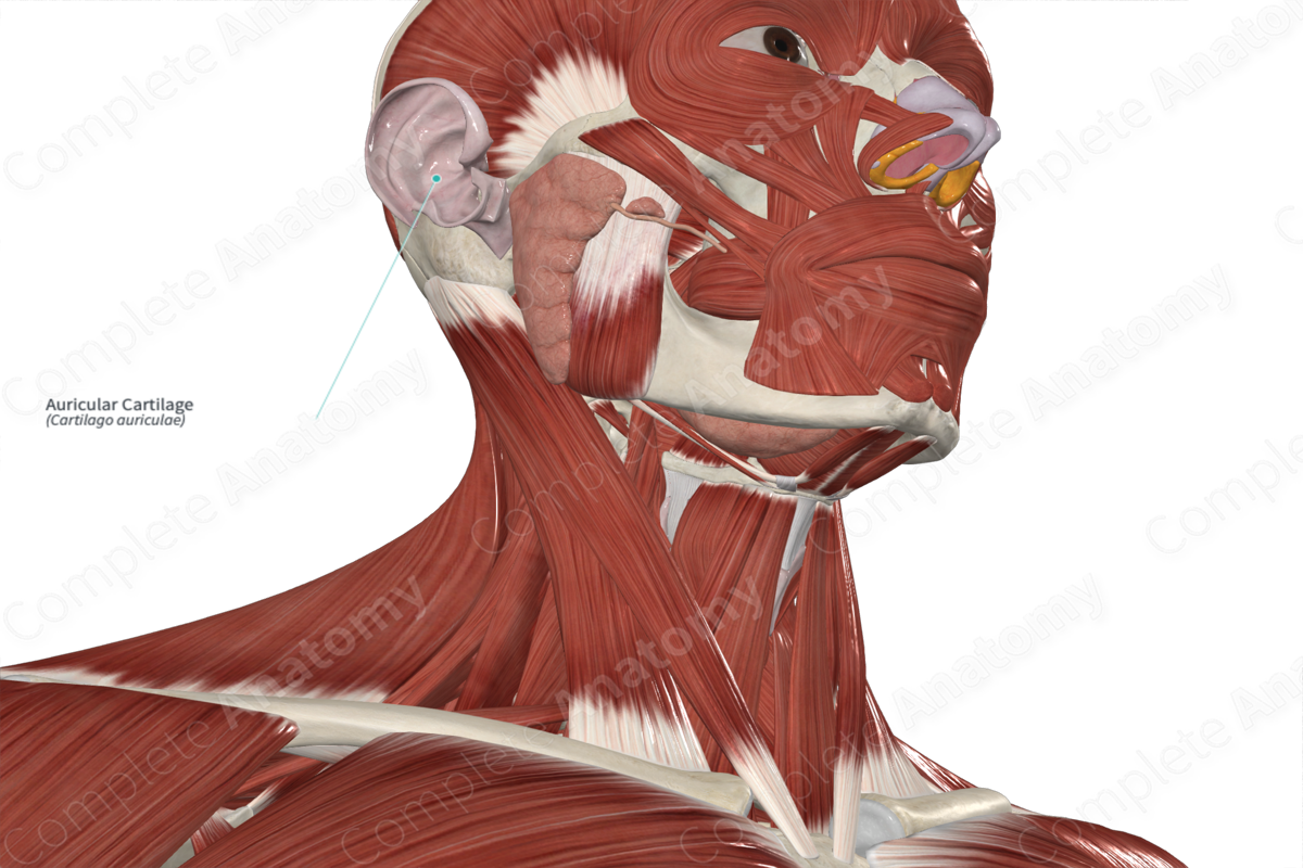
The external ear is composed of both the auricle and the external acoustic meatus. The auricle is composed of a single plate of elastic fibrocartilage, covered by thin skin. The auricle is formed by the auricular cartilage with two exceptions. At the lobule (or earlobe) and the area between the tragus and the crus of the helix, there is no cartilage beneath the skin.
The auricular cartilage is primarily composed of the helix, the elevated outer margin, and the concha, the hollow next to the ear canal. Other cartilaginous features are the tragus, found anterior to the opening of the external acoustic meatus, the antitragus, located opposite the tragus, and the antihelix, a smaller rim parallel to the helix.
Related parts of the anatomy
Anatomical Relations
The external ear projects from the side of the head by variable amounts. The cartilaginous auricle is connected to the surrounding structures by ligaments and muscles and is continuous with the external acoustic meatus.
Function
The auricle conducts sound waves from the external environment through the external acoustic meatus towards the tympanic membrane.
List of Clinical Correlates
—Auricular hematoma
Learn more about this topic from other Elsevier products
Auricular Cartilage

Auricular cartilage is a single plate of elastic cartilage normally containing perforations through which blood vessels pass between the convex and concave surfaces of the pinnae.


