Connective Tissue of Orbital Cavity (Left)
Textus connectivus cavitatis orbitalis
Read moreDescription
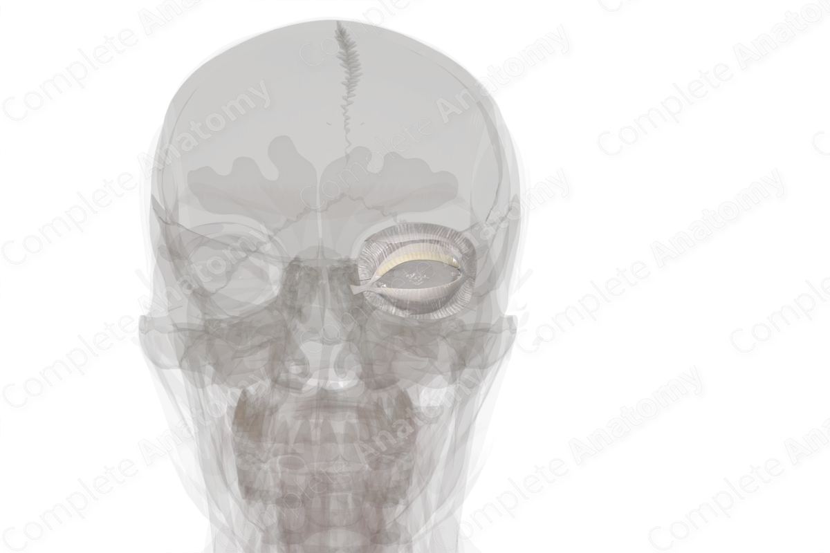
The connective tissues in and around the orbit all function to support the structures within the orbit. They are imperative to the functioning of the extraocular muscles and maintaining a normal location of the eyeball, as well as some protective functions.
There are a number of connective tissues located within the orbit, ranging from loose connective tissues like the orbital septum, to the fibrous superior and inferior tarsal plates made of dense connective tissue.
The contents of the orbit and their movements are minuscule and particular, so it is important that the associated supportive and protective connective tissues are functioning correctly. In conditions such as systemic sclerosis, the connective tissue stiffens throughout the body. When this affects the orbit, it can lead to eyelid stiffness and dry eye syndrome.


Related parts of the anatomy
Learn more about this topic from other Elsevier products

