Inferior Retrodiscal Lamina of Temporomandibular Joint (Right)
Lamina retrodiscus inferior articulationis temporomandibularis
Read moreStructure
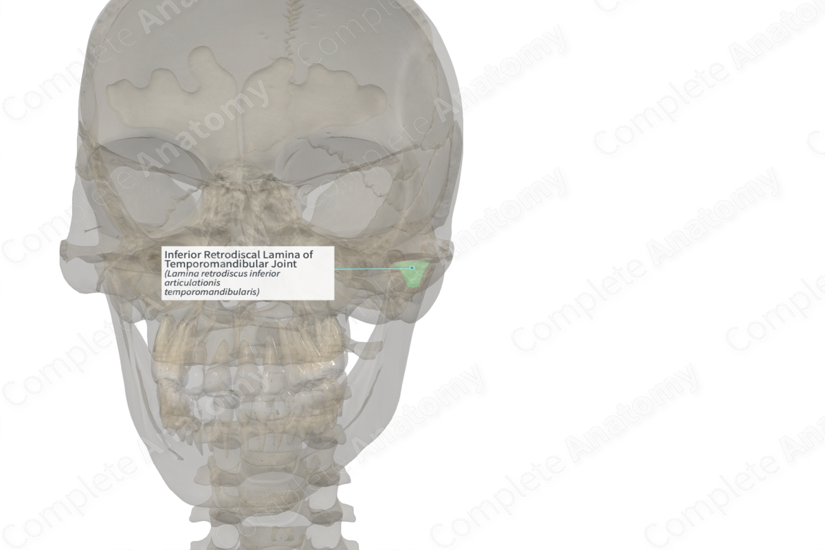
The inferior retrodiscal lamina is composed of collagenous tissue.
Anatomical Relations
The inferior retrodiscal lamina attaches to the posteroinferior part of the articular disc. It extends to the posterior aspect of the mandibular condyle (Miloro & Peterson, 2012).
Function
The inferior retrodiscal lamina is not elastic like the superior lamina, therefore, acts to maintain a strong relationship to the mandibular condyle.
List of Clinical Correlates
—Temporomandibular disorder
References
Miloro, M. & Peterson, L. J. (2012) Peterson's Principles of Oral and Maxillofacial Surgery. People's Medical Publishing House-USA.
Learn more about this topic from other Elsevier products
Temporomandibular Joint

The temporomandibular joint (TMJ) is a bilateral synovial joint between the skull and the mandible comprising the glenoid fossa of the temporal bone, the condylar head of the mandible, and the articular cartilage and disc.
