Structure
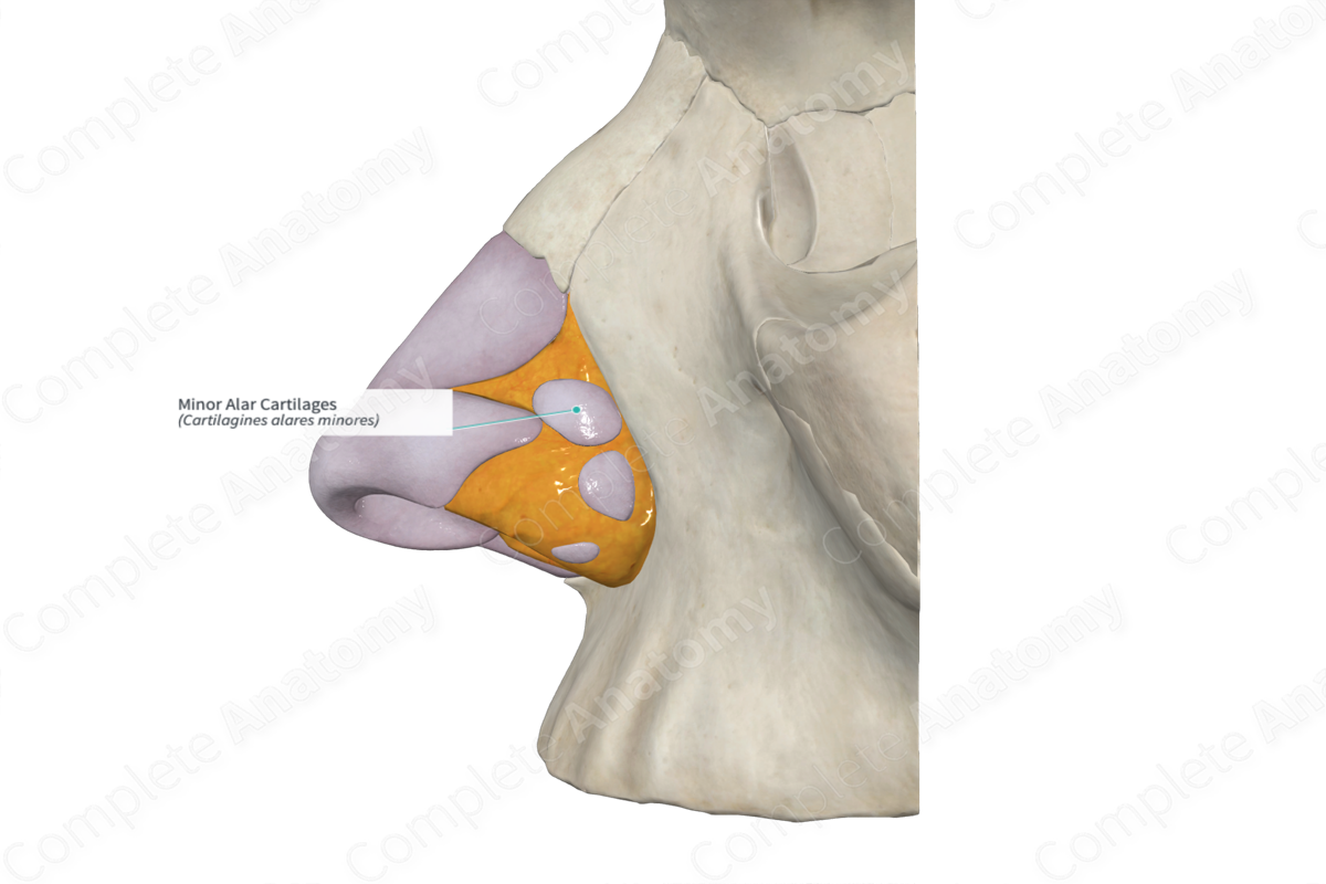
The minor alar cartilages are paired multiple plates of cartilage forming the posterior and lateral cartilaginous skeleton of the external nose.
There are typically three or four ‘islands’ of minor alar cartilages embedded in alar fibrofatty tissue. They complete the posteroinferior lateral wall of the external nose where they attach to the frontal process of the maxilla bone, thereby attaching the external nose to the facial skeleton.


Related parts of the anatomy
Anatomical Relations
The minor alar cartilages are found in the fibrous membrane between the lateral border of the major alar cartilage and the frontal process of the maxilla.
Function
The minor alar cartilages are the fibrofatty alar tissue contributes to the posterolateral rim of the nares, giving them flexibility.
Learn more about this topic from other Elsevier products
Nasal Cartilages

Intercrural ligament (suspensory ligament) is a ligamentous connection between the cephalic border of entire alar cartilages.
