Superior Retrodiscal Lamina of Temporomandibular Joint (Left)
Lamina retrodiscus superior articulationis temporomandibularis
Read moreStructure
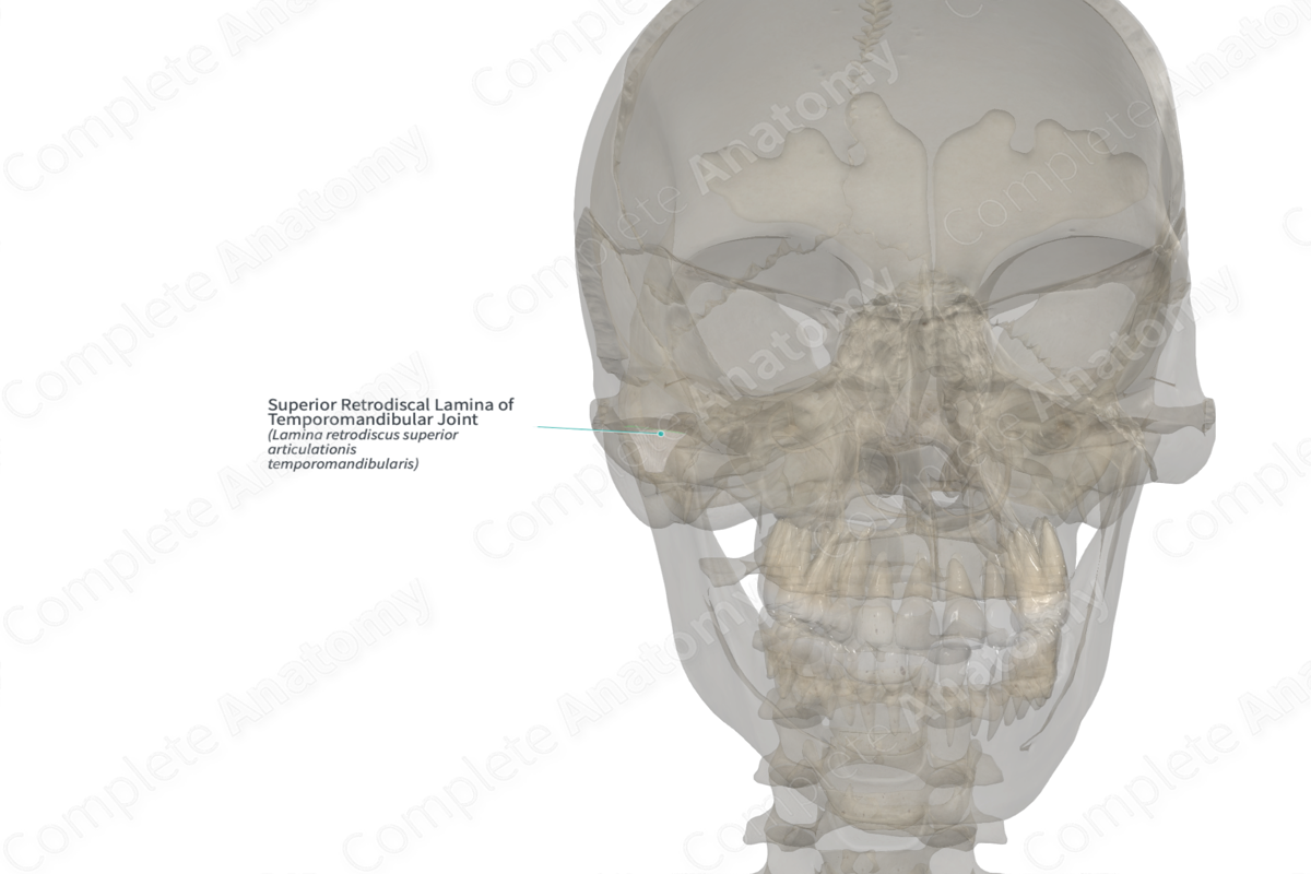
The superior retrodiscal lamina consists of connective tissue with many elastic fibers.
Anatomical Relations
The superior retrodiscal lamina attaches to the posterosuperior part of the articular disc. It extends to the posterior wall of the mandibular fossa (the petrotympanic fissure) (Miloro & Peterson, 2012).
Function
The superior retrodiscal lamina allows anterior translation of the articular disc over the articular eminence.
List of Clinical Correlates
—Anterior disc displacement
—Temporomandibular disorder
References
Miloro, M. & Peterson, L. J. (2012) Peterson's Principles of Oral and Maxillofacial Surgery. People's Medical Publishing House-USA.
Learn more about this topic from other Elsevier products
Temporomandibular Joint

The temporomandibular joint (TMJ) is a bilateral synovial joint between the skull and the mandible comprising the glenoid fossa of the temporal bone, the condylar head of the mandible, and the articular cartilage and disc.
