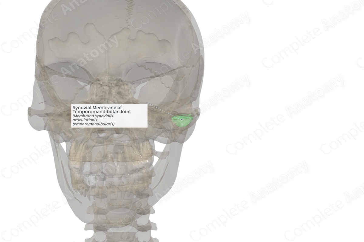
Synovial Membrane of Temporomandibular Joint (Left)
Membrana synovialis articulationis temporomandibularis
Read moreStructure
The synovial membrane of the temporomandibular joint is the inner layer of the articular capsule of the temporomandibular joint. It is composed of loose connective tissue with a smooth inner surface which lines the joint cavity. It may be broken up into superior and inferior parts, which lines the joint cavity above and below the articular disc of the temporomandibular joint, respectively.

Related parts of the anatomy
Anatomical Relations
The blood supply to the synovial membrane of the temporomandibular joint is via the superficial temporal artery.
Function
The synovial membrane produces synovial fluid that fills both the upper and lower compartments of the temporomandibular joint. This fluid acts to lubricate the joint. The intermediate portion at the center of the articular disc is avascular and, therefore, gains its nutrients from the synovial fluid.
List of Clinical Correlates
—Synovitis
Learn more about this topic from other Elsevier products

