Articular Capsule of Hip Joint (Left)
Capsula articularis articulationis coxae
Read moreStructure
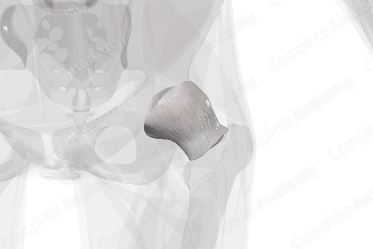
The articular capsule of the hip joint is a sac-like structure that enclosed the hip joint. It is composed of two layers. The external layer, the fibrous membrane, is composed of strong, dense fibrous tissue. The internal layer, the synovial membrane, lines the joint cavity and is composed of loose connective tissue. The anterosuperior aspect of the articular capsule is thicker because that's the point where maximal stress takes place.


Related parts of the anatomy
Anatomical Relations
The articular capsule of the hip joint is attached to the acetabular margin, the acetabular labrum, the transverse acetabular ligament, and the obturator foramen. From the capsule’s acetabular attachment point, it projects laterally to encircle the head and neck of the femur, and then attaches to the intertrochanteric line, the base of the femoral neck, intertrochanteric crest, and at a point near the lesser trochanter.
The attachment point of the capsule to the femur is situated distal to the growth plate of the head of the femur, making the femur’s upper epiphysis completely intracapsular. The joint capsule lies deep to muscles and tendons, and is separated anteriorly from the psoas major and iliacus muscles by the iliopectineal bursa. The joint capsule is reinforced by the iliofemoral, pubofemoral, and ischiofemoral ligaments.
Function
The joint capsule of the hip joint ensures that the joint is sealed, thus, keeping the lubricating synovial fluid within the joint. It provides passive stability to the joint by limiting the joint movement. Additionally, it provides active stability by containing numerous proprioceptive nerve endings which relay mechanical information back to the central nervous system (Ralphs and Benjamin, 1994).
List of Clinical Correlates
—Rheumatoid arthritis
—Osteoarthritis
—Capsule laxity and microinstability
References
Ralphs, J. R. and Benjamin, M. (1994) 'The joint capsule: structure, composition, ageing and disease', Journal of Anatomy, 184 (Pt 3), pp. 503-509.
Learn more about this topic from other Elsevier products
Hip Joint

The hip joint is a synovial articulation between the acetabulum of the pelvis and the proximal femur.
