Description
The articulating surfaces of the knee joint include the condyles of the femur, which articulate with the proximal flat tibial condylar surface, called the tibial plateau. Like other synovial joints, the articulating surfaces in the knee joint are covered by hyaline cartilage. It provides a smooth surface to reduce friction, thus allowing the articulating bones to slide freely against each other.
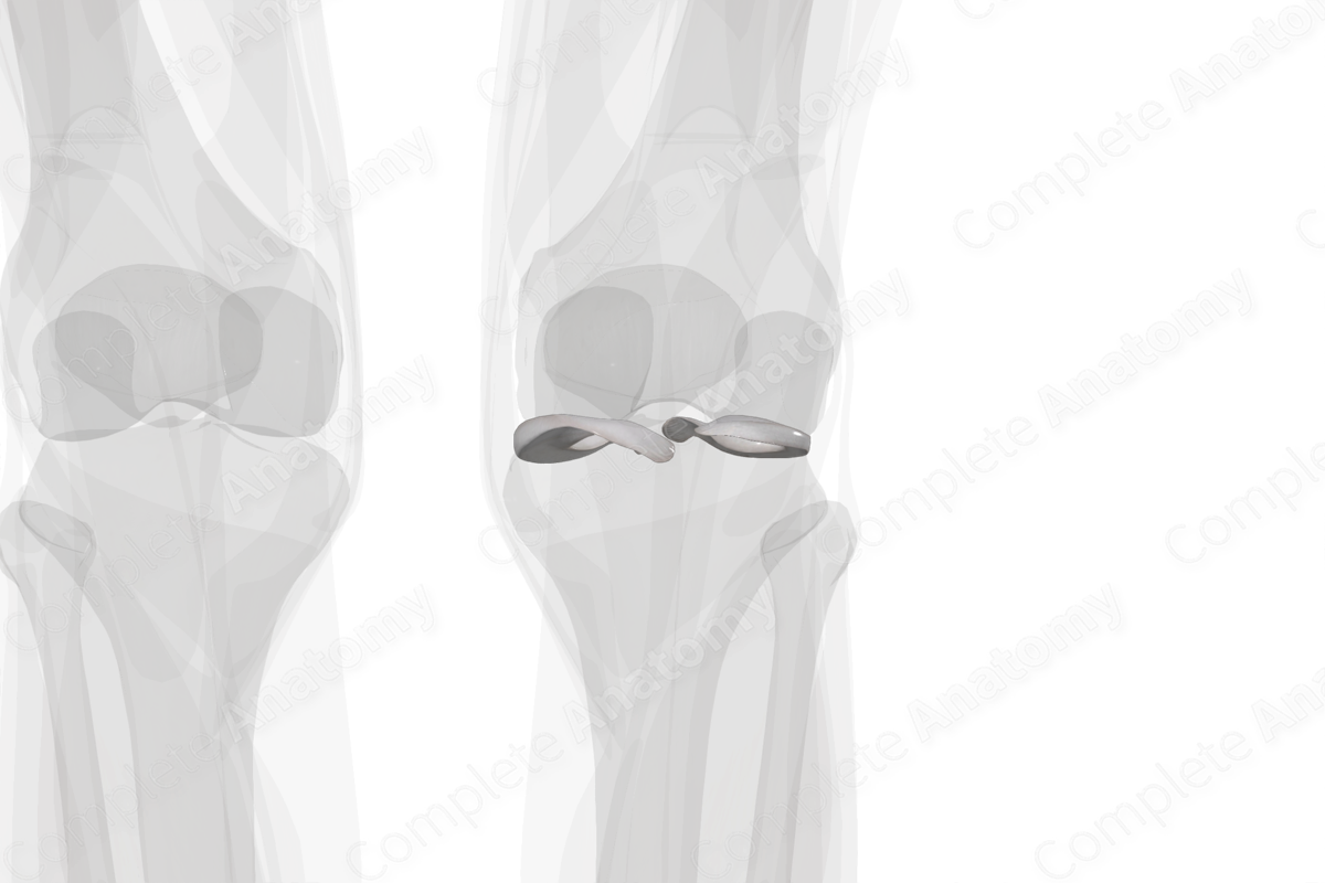
Between the ends of the femoral condyles and the tibial plateau, lie two crescent-shaped, fibrocartilaginous structures called the menisci. The menisci function to widen and deepen the tibial articular surfaces to receive the femoral condyles, thus providing stability to the knee joint. Both the lateral and medial menisci slide backwards with flexion and forwards in extension at the knee joint and act as shock absorbers in the knee joint.
The menisci may be injured or torn by twisting the knee or applying direct force to them, as seen in contact sports. The lateral meniscus moves more compared to the medial meniscus, and hence is less prone to injury compared to the medial meniscus. The latter is denied free mobilization because of its additional attachments to the fibrous capsule and the medial collateral ligament.


Related parts of the anatomy
List of Clinical Correlates
—Meniscal tears
—Unhappy triad
Learn more about this topic from other Elsevier products

